
Three destructive processes converge in Alzheimer disease. Amyloid accumulates outside neurons. Tau collapses the transport system inside them. Amyloid also weakens cerebral vessel walls. Together these processes explain why the disease begins in memory circuits and spreads in a predictable sequence.
Amyloid plaques: the extracellular cascade
The amyloid-beta 42 peptide, abbreviated Aβ42, is cleaved from amyloid precursor protein (APP). Cleavage happens in two steps, first by β-secretase and then by γ-secretase. Aβ42 is insoluble, so single molecules clump into small clusters called oligomers. Oligomers then pack into the dense plaques that sit between neurons.
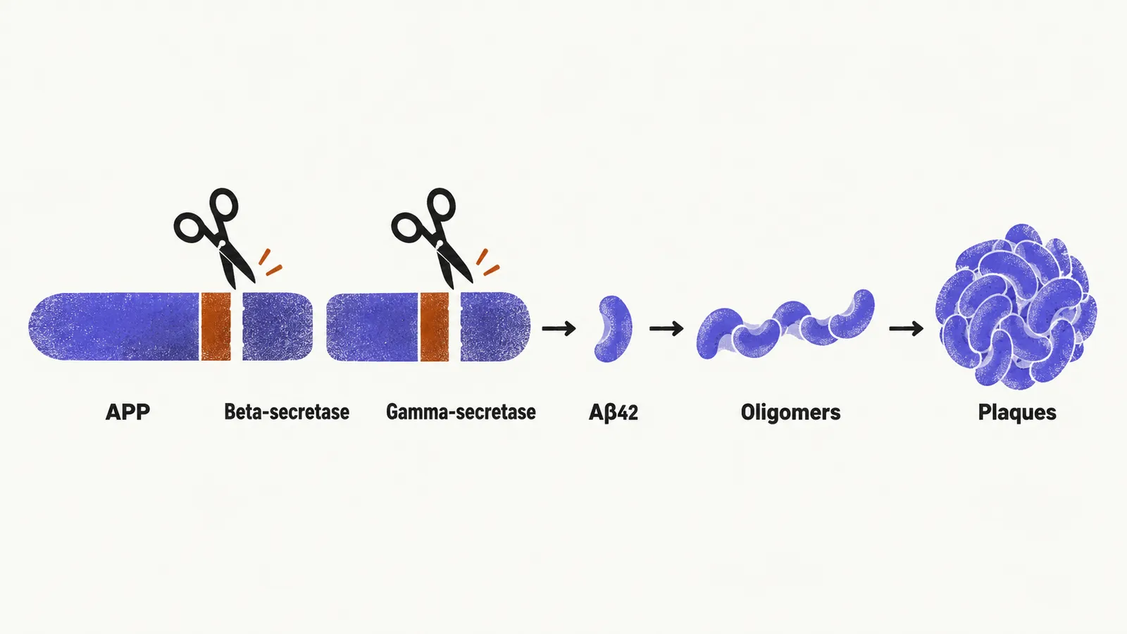
Oligomers may injure synapses more than plaques do. They disturb synaptic plasticity, the process by which neurons strengthen or weaken connections. Memory networks begin to fail before neurons die. Plaques also provoke a chronic microglial inflammatory response. That inflammation damages surrounding healthy tissue.
Amyloid deposition can begin a decade or more before any symptom. During this preclinical stage the patient stays cognitively normal while pathology accumulates. Measuring Aβ42 in cerebrospinal fluid (CSF) and amyloid PET (positron emission tomography) can detect disease before bedside testing can.
Neurofibrillary tangles: the intracellular collapse
Tau protein normally stabilises microtubules. Microtubules are the tracks that carry organelles and synaptic cargo down the axon. In Alzheimer disease tau becomes hyperphosphorylated. It detaches from the microtubules and aggregates into paired helical filaments. These filaments twist into neurofibrillary tangles inside the neuron.
When tau releases the microtubules, axonal transport collapses. The synapse loses its supply chain and weakens. The neuron then dies. Tangle density tracks cognitive decline more closely than plaque burden does. Plaques set the disease in motion. Tangles destroy neurons and function.
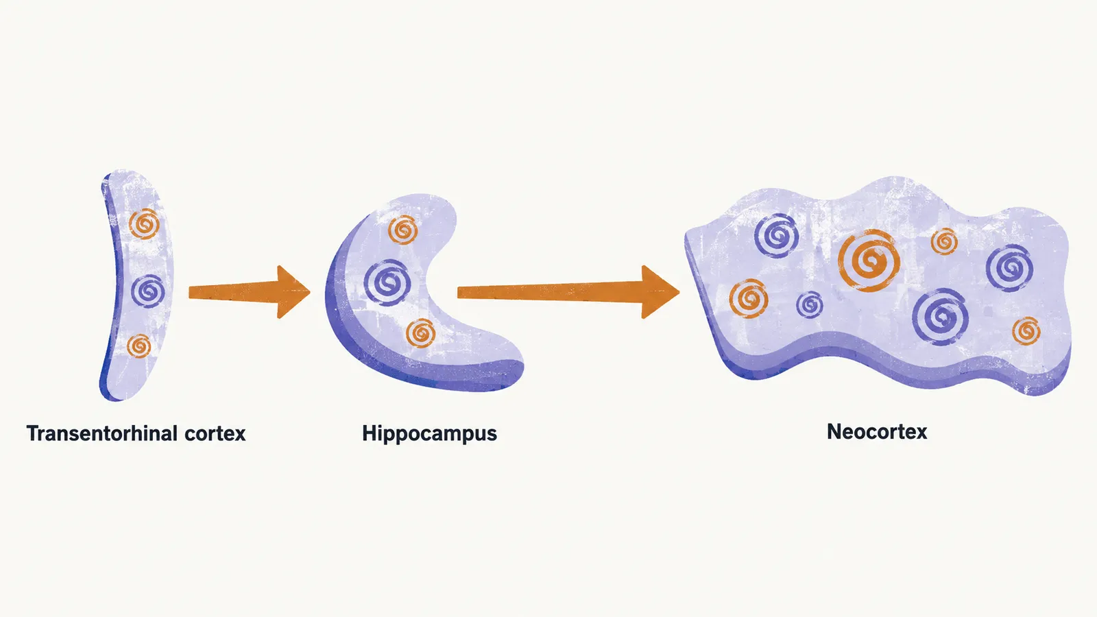
Tangle pathology spreads in a fixed sequence. It begins in the transentorhinal cortex. It then reaches the hippocampus. It finally reaches the neocortex. This sequence mirrors the clinical course. Memory fails first, then language and visuospatial function, and finally executive function and behaviour.
Cerebral amyloid angiopathy: the vascular dimension
Amyloid-beta does not stay only between neurons. It also deposits inside cerebral vessel walls, which is called cerebral amyloid angiopathy, abbreviated CAA. Infiltrated walls turn brittle and prone to rupture. Rupture produces haemorrhage at the convexity of the lobes, called lobar haemorrhage. This site differs from the deep bleeds of hypertensive small-vessel disease. Microbleeds on MRI gradient-echo sequences reflect the same vessel process. Extensive CAA raises the risk of major bleeding, so antiplatelet and anticoagulant decisions need added care in these patients.
Macroscopic pathology: selective atrophy
Neuronal loss and reactive gliosis, the proliferation of supporting glial cells around damaged tissue, shrink the brain. Shrinkage concentrates in the temporal and parietal lobes. Ventricular enlargement and widened sulci accompany the atrophy.
Early hippocampal destruction explains the cardinal symptom. The hippocampus gates new memories into long-term storage, so recent memory fails while old memories persist. Pure Alzheimer disease shows no focal vascular lesions. That absence helps distinguish it from vascular dementia on structural MRI.
Neurotransmitter deficits: why the drugs work the way they do
Beyond structural loss, specific transmitter systems fail, and current drugs act on them. The nucleus basalis of Meynert is a cholinergic cluster in the basal forebrain, meaning its neurons use acetylcholine as their transmitter. It projects to the hippocampus and cortex. It degenerates early in the disease. The resulting cholinergic deficit worsens attention, learning, and memory. Acetylcholinesterase inhibitors slow acetylcholine breakdown. They amplify what surviving neurons still release. The effect is symptomatic rather than disease-modifying.
A second transmitter system is glutamate, the main excitatory transmitter. Excess glutamate signalling at NMDA receptors admits toxic amounts of calcium into neurons. Calcium activates destructive intracellular enzymes, a process called excitotoxicity. Memantine is an NMDA receptor antagonist. It dampens this cascade without altering the underlying degeneration.
Serotonergic and noradrenergic systems matter for mood and behaviour. Declining serotonergic and noradrenergic transmission contributes to apathy, depression, and agitation. These symptoms reflect direct circuit failure rather than reaction to cognitive loss.
Risk factors
Risk factors fall into age, genetics, and the brain’s capacity to compensate. Advanced age is the strongest known risk factor: risk rises steeply with age. Women are affected more often than men, partly because they live longer on average. Reported prevalence figures vary across populations because case ascertainment and methodology differ.
Most cases are sporadic and late-onset. A small minority are familial and early-onset. These cases appear from the sixth decade or earlier. They link to mutations in APP or in the presenilin genes PSEN1 and PSEN2. Presenilin genes encode parts of the γ-secretase complex, the enzyme that cleaves Aβ42 from APP. The mutations shift production toward aggregation-prone Aβ42.
In the common late-onset form, APOE ε4 is the strongest genetic risk factor. Risk rises with each ε4 copy. The ε2 isoform carries lower risk. APOE ε4 appears to impair amyloid clearance and promote deposition. The mechanism remains debated.
Cognitive reserve means denser networks built through education and lifelong mental stimulation. Reserve delays clinical onset without slowing pathological accumulation, so the brain compensates longer before symptoms surface.
Two further factors carry added risk: head trauma and a first-degree family history.