
The hematoma after intracerebral hemorrhage, the collection of blood within the brain, is not static. Expansion, meaning growth of that collection, occurs early, most often within the first 2 hours, and predicts worse outcome. Everything in this note serves that clock: predict who expands, reverse what drives bleeding, and hold pressure steady.
Predicting expansion
Neuroimaging markers, together with time since onset and antithrombotic use, help predict expansion risk. The CTA spot sign, seen on CT angiography, is focal pooling of contrast within the hematoma, marking active extravasation, or leakage of blood from a vessel. It significantly increases the likelihood of hematoma growth. About 30% of patients scanned within 6 hours of onset show it.
Reversing anticoagulation
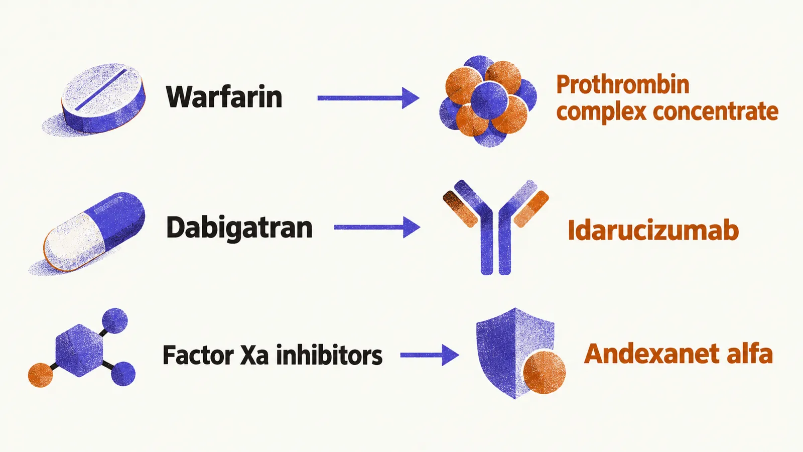
Reversal means neutralizing an anticoagulant’s effect, and acute reversal pairs agent to anticoagulant. Prothrombin complex concentrate reverses warfarin-type vitamin K antagonists. Idarucizumab reverses the thrombin inhibitor dabigatran. Andexanet alfa reverses factor-Xa inhibitors such as rivaroxaban, apixaban, and edoxaban. Availability and cost vary by setting, so the pairing is the teaching point rather than any local protocol.
Blood pressure, what to avoid, and cerebellar evacuation
Acute blood-pressure lowering after mild-to-moderate hemorrhage aims for smooth, sustained control with limited variability. That pattern appears to reduce expansion and improve outcome.
Some treatments do not help. Prophylactic corticosteroids and continuous hyperosmolar therapy show no outcome benefit. Platelet transfusion outside emergency surgery or severe thrombocytopenia appears to worsen outcome.
Surgery is a separate decision from these medical measures. For cerebellar hemorrhage, immediate evacuation with or without external ventricular drain now includes volume above 15 mL alongside deterioration, brainstem compression, and hydrocephalus.
Bleeding does not always enter brain tissue; it can also spread over the brain surface, which is the territory of subarachnoid haemorrhage.