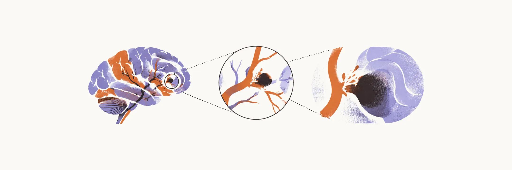
Intracerebral haemorrhage is bleeding directly into brain tissue, most often from rupture of small deep penetrating arteries damaged by chronic hypertension. It accounts for roughly 10–15% of strokes yet carries the highest reported mortality among stroke subtypes, around 60% at one year. The blood itself causes the damage: the hematoma, the collected blood, compresses adjacent tissue as a mass, raises intracranial pressure, and provokes edema and secondary ischemia around it.
Chronic hypertension injures the lenticulostriate and other small penetrating end-arteries through lipohyalinosis, a degenerative weakening of the wall under pulsatile stress. Without collateral supply, rupture lets blood dissect into tissue under arterial pressure over minutes to hours. The basal ganglia (especially the putamen), thalamus, pons, and cerebellum are the typical sites because those territories depend on these vulnerable vessels. Bleeding in an atypical lobar location, within a cerebral lobe rather than a deep structure, in an older adult should instead prompt consideration of amyloid angiopathy, arteriovenous malformation, tumor, or infection. Anticoagulant use, vasculitis, and cocaine are additional causes.
Presentation by location
The hallmark is a stroke that worsens: sudden severe headache, vomiting from raised pressure, and progressive deterioration that may reach coma, as the hematoma expands. Location shapes the syndrome. Putaminal bleeding resembles a middle cerebral artery stroke with contralateral weakness and sensory loss but adds headache, vomiting, and progression. Thalamic bleeding produces contralateral sensory loss predominating over weakness. Pontine bleeding can produce locked-in syndrome, with intact consciousness but quadriplegia and facial paralysis sparing vertical eye movements, because the tegmentum supporting arousal is spared while the motor basis is destroyed. Cerebellar bleeding presents with ataxia, vertigo, and occipital headache and can compress the brainstem quickly because the posterior fossa has little spare room.
Diagnosis on CT
Non-contrast CT is immediate and usually definitive. Acute blood appears hyperdense, bright white against brain tissue, from the moment of bleeding, unlike ischemia which is often invisible for hours. CT also shows hematoma size and location, ventricular extension that worsens prognosis through hydrocephalus, and midline shift or early herniation. Blood-sensitive MRI sequences help when a lobar pattern suggests amyloid angiopathy with its characteristic microbleeds.
Blood pressure and surgery
Blood pressure control is the first priority because the hematoma remains under arterial pressure and hypertension drives expansion. Lowering aims for smooth, sustained control with limited variability.
Surgical evacuation is selective because routine surgery has poor supporting evidence. Cerebellar hemorrhage with volume above 15 mL calls for immediate evacuation, alongside deterioration, brainstem compression, and hydrocephalus, with or without external ventricular drainage. Hemispheric hematoma larger than roughly 50 cubic cm with clinical deterioration is the other commonly cited indication. Surgery is generally not pursued in deep coma, where underlying injury is usually too extensive to change outcome.
Measures to avoid
Several measures are avoided. Osmotic agents such as mannitol or glycerol are reserved for rising pressure, herniation, or deterioration rather than given prophylactically, and patients are kept still because straining, agitation, and vomiting raise intracranial pressure further. Corticosteroids are not recommended for the edema of intracerebral haemorrhage. They help vasogenic edema around tumors but have not shown benefit for hemorrhagic compressive and cytotoxic edema and may worsen outcome through hyperglycemia and immunosuppression. Platelet transfusion outside emergency surgery or severe thrombocytopenia appears to worsen outcome rather than help.
Prognosis and early expansion
Prognosis tracks hematoma volume, location, consciousness at presentation, and ventricular extension. Survivors often carry substantial lasting disability.
Expansion of the hematoma occurs early, most often within the first 2 hours, and predicts worse outcome. Expansion timing, reversal pairings, and the imaging marker that predicts growth are set out under expansion and reversal.