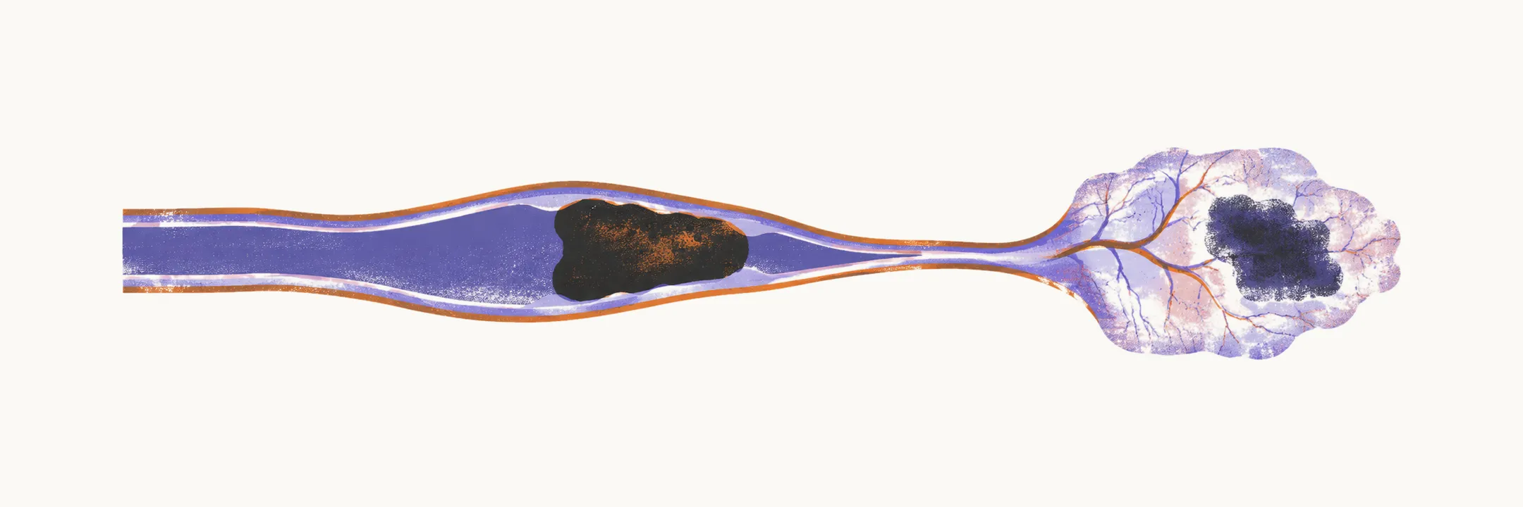
Ischemic stroke is occlusion of a cerebral artery by thrombus (a clot formed in the vessel) or embolus (material carried there from elsewhere), depriving downstream tissue of blood and oxygen. It accounts for roughly 85% of strokes. The central tension is between dead tissue and tissue at risk: an irreversibly damaged core surrounded by a rim of hypoperfused (under-supplied) but still viable tissue called the ischemic penumbra. Acute treatment exists to rescue that rim before it collapses into necrosis, meaning cell death.
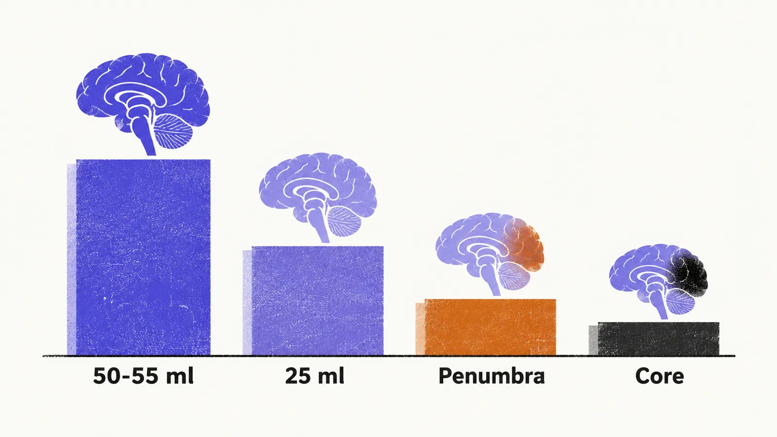
Normal cerebral blood flow runs near 50–55 ml per 100 g per minute. Around 25 ml per 100 g per minute, electrical activity slows and membrane pumps begin to fail, leaving cells dysfunctional but potentially recoverable. Continued ischemia leads to cytotoxic edema (swelling of injured cells), calcium influx, and excitotoxic injury at the core. Symptoms that evolve over hours often reflect penumbra converting to infarct, meaning the area of dead tissue. That is why reperfusion is organized around speed and why imaging of the core-penumbra mismatch guides treatment selection.
How the artery becomes occluded
The blocking clot either forms in the artery itself or arrives from a source elsewhere, and a smaller group of causes lies in the arterial wall or the blood. These routes define thrombotic, embolic, and other mechanisms.
Thrombotic stroke, roughly half of ischemic events, arises when atherosclerotic plaque narrows a vessel, often at the carotid bifurcation or within the Circle of Willis where turbulent flow promotes plaque, until thrombosis completes the occlusion. It predominates in older patients with chronic vascular risk factors.
Embolic stroke, roughly one quarter, occurs when clot formed elsewhere lodges in a cerebral artery. The heart is the usual source, and atrial fibrillation is the single most common cardiac cause: stagnant blood in the fibrillating atrium clots, and the clot later ejects into the cerebral circulation. Other sources include endocarditis, mural thrombus after myocardial infarction, valvular disease, paradoxical embolism through a patent foramen ovale, and aortic arch atheroma. Rare causes include gas, fat, or tumor emboli.
Other causes include arterial dissection after trauma or neck injury, cerebral vasculitis with irregular narrowing on angiography, and hypercoagulable states. When no cardiac or arterial source is found, carotid, aortic arch, and cardiac imaging become the next steps.
Localizing the occlusion
Each artery supplies a recognizable territory, so the deficit pattern points to the occluded vessel. Lateral convexity syndromes, which involve the outer surface of the hemisphere, with language impairment or neglect of one side of space point anteriorly. Isolated hemianopia, loss of half of the visual field, points posteriorly, and crossed patterns of cranial-nerve signs on one side with body signs on the other point to the brainstem. The full artery-to-deficit patterns are set out under territory syndromes.
Presentation and first imaging
Ischemic stroke presents as a sudden focal deficit arriving over seconds to minutes. Headache is characteristically absent; prominent headache, vomiting, or rapid deterioration instead favors haemorrhage. Early weakness may appear flaccid with reduced reflexes before upper motor neuron signs emerge over days.
The first study is non-contrast CT, performed urgently, chosen to exclude blood rather than to show early ischemia, which is often invisible for hours. Early ischemic signs, when present, include a hyperdense vessel, loss of gray-white distinction such as the insular ribbon, and sulcal effacement. Diffusion-weighted MRI detects established injury earlier, and perfusion imaging maps tissue at risk. Follow-up vascular and cardiac imaging then seeks the source and guides prevention.
Duration separates completed stroke from a transient ischemic attack. A deficit lasting less than 24 hours, usually minutes to 2 hours, is classified as a transient ischemic attack rather than completed stroke. The mechanism is the same; reperfusion simply arrived before permanent damage.