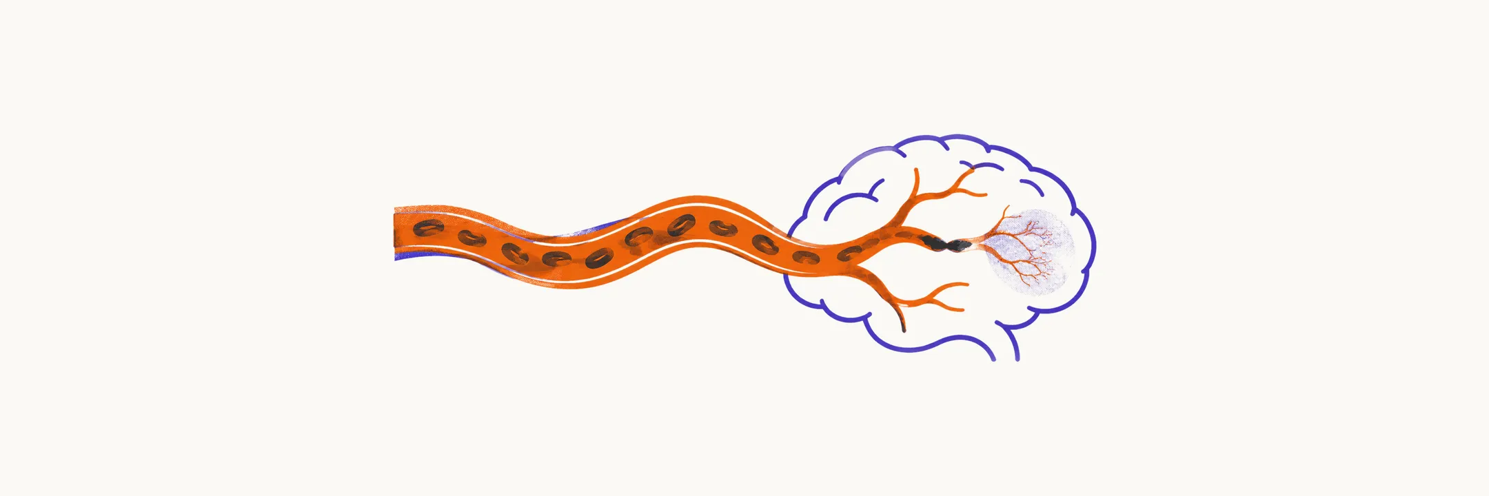
Bedside localization starts from one rule. Each artery supplies a recognizable territory, so the deficit pattern points to the occluded vessel. Anterior-circulation strokes from carotid flow into the anterior and middle cerebral arteries account for about 70% of ischemic strokes. Throughout, contralateral means on the opposite side of the body from the lesion and ipsilateral means on the same side.
Middle cerebral artery
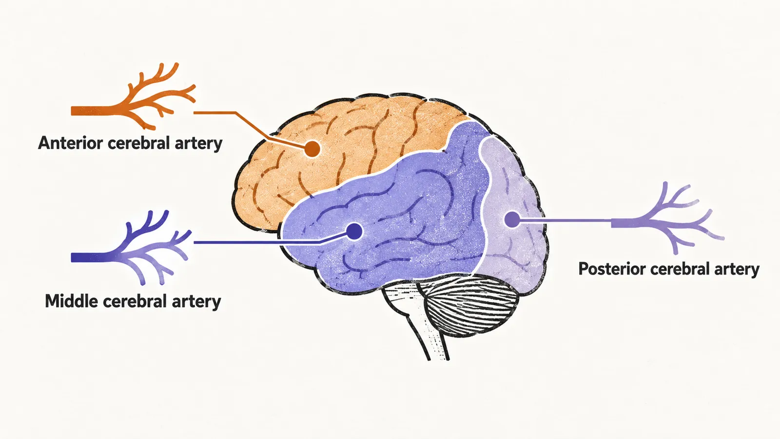
The middle cerebral artery supplies most of the lateral convexity, including the face and arm areas of the motor and sensory maps, the dominant-hemisphere language areas, and the optic radiations. Occlusion is highly symptomatic. It typically produces contralateral weakness and sensory loss most marked in the face and arm, contralateral homonymous hemianopia (loss of the same half of the visual field in both eyes), and gaze deviation toward the lesion from frontal eye field involvement. Left-sided occlusion may add Broca or Wernicke aphasia, the two language disorders. Right-sided occlusion may add anosognosia (unawareness of the deficit) and hemispatial neglect (failure to attend to one side of space).
Anterior cerebral artery
The anterior cerebral artery supplies the medial frontal and superior parietal lobes, including the leg representation. It accounts for roughly 20% of ischemic strokes. Occlusion produces contralateral leg-predominant weakness, sometimes with executive or behavioral change, and usually spares language. Presentation is often atypical or subtle, with abulia (reduced initiative and motivation) and alien-hand phenomena as signature non-motor features. That subtlety causes underrecognition and delayed treatment.
Posterior cerebral artery
The posterior cerebral artery branches supply the midbrain, thalamus, and the temporal, occipital, and occipitoparietal cortices. Occlusion produces contralateral homonymous hemianopia with macular sparing, meaning central vision is preserved, with motor function typically preserved. That visual field pattern is the anchor bedside sign for posterior territory. Left-sided lesions can rarely produce alexia without agraphia, inability to read with preserved writing, where visual input cannot reach the language area.
Brainstem: the crossed pattern
Brainstem infarction produces a crossed pattern: cranial nerve signs on the same side as the lesion with body weakness or sensory loss on the opposite side. Two syndromes organize the possibilities. Weber syndrome of the ventral midbrain combines an ipsilateral third nerve palsy (ptosis, down-and-out eye, dilated pupil) with contralateral spastic hemiparesis, from paramedian posterior cerebral or basilar-tip perforator ischemia. Wallenberg syndrome of the lateral medulla is the most common vascular brainstem syndrome, usually from vertebral-artery perforator or posterior inferior cerebellar artery occlusion. It combines ipsilateral Horner syndrome, facial pain and temperature loss, vertigo, dysphagia, and hoarseness with contralateral body pain and temperature loss through the spinothalamic tract. Strength is typically preserved because the pyramidal tract runs medially and is spared. Unlike medial medullary syndrome, Wallenberg syndrome produces no tongue deviation and no contralateral hemiparesis.
| Feature | Weber (midbrain) | Wallenberg (lateral medulla) |
|---|---|---|
| Artery | Paramedian posterior cerebral or basilar-tip branches | Posterior inferior cerebellar or vertebral artery |
| Same-side signs | Third nerve palsy | Horner syndrome, facial pain and temperature loss, vertigo, dysphagia |
| Opposite-side signs | Hemiparesis | Body pain and temperature loss |
| Strength | Reduced | Usually preserved |
Naming the occluded vessel is the first step. Whether that vessel can be reopened depends on timing and on whether the patient is eligible for reperfusion, which is the question acute management addresses.