
Because a raised prolactin level has many causes, the diagnosis is built by excluding the common ones first and then using the prolactin level and imaging together to decide whether a tumour is present. Each of those two pieces of information can mislead on its own, so they are read against each other.
Ruling out other causes
In patients with hyperprolactinemia it is mandatory first to rule out the other causes of hyperprolactinemia, which are:
- pregnancy
- lactation
- hypothyroidism
- drug-induced hyperprolactinemia
- renal failure
- liver failure
- hypothalamic defects
These are common and reversible, so they come before any imaging. Hypothyroidism raises prolactin by increasing TRH, which stimulates the lactotrophs; renal and liver failure reduce the clearance of prolactin; and drugs remove part of the dopaminergic brake on the lactotrophs.
What remains after exclusion
After ruling out the causes above, three possibilities will remain, which are:
- microadenoma, which is 4 to 5 folds more prevalent than macroadenoma
- macroadenoma
- no visible tumour at all; at this stage these patients are considered to have idiopathic hyperprolactinemia
Idiopathic hyperprolactinemia may simply be a microadenoma too small to be seen, and it usually remains stable on follow-up.
Imaging
MRI is the modality of choice when a prolactinoma is suspected, using T2-weighted coronal sections and T1-weighted coronal sections before and after gadolinium (the contrast agent). On T1-weighted MRI, a prolactinoma is usually hypo-intense or iso-intense, and rarely hyper-intense, which happens in the case of haemorrhagic complications of prolactinoma. On T2-weighted MRI, usually 80% of prolactinomas are hyper-intense.
CT scan would be helpful in the case of macroprolactinoma with suspected invasion of the bony structures, which can present with CSF (cerebrospinal fluid) rhinorrhea; this happens mostly in men with macroprolactinoma after shrinkage of the mass after dopamine agonist therapy.
Prolactin level and tumour size
Once imaging has found an adenoma, its size is read against the prolactin level. If the adenoma which is found is smaller than 10 mm it is considered as microadenoma, and if it is bigger than 10 mm it is considered as macroadenoma. Usually in patients with microadenoma the serum level of prolactin is < 200 ng/ml, and in patients with macroadenoma the level of serum prolactin is mostly greater than 200 ng/ml, and mostly it is more than 500 ng/ml.
When the level and the tumour size do not match
Sometimes the prolactin level is not correlated with the size of a tumour that imaging shows to be a macroprolactinoma, for instance a macroprolactinoma with hyperprolactinemia of 100-200 ng/ml. Two different conditions then have to be differentiated during the diagnostic panel:
- macroprolactinoma with hyperprolactinemia correlated with the size of the tumour
- macroprolactinoma with hyperprolactinemia not correlated with the size of the tumour; in this case the tumour is not a functioning tumour that can release prolactin into the blood, and the cause of the hyperprolactinemia is only compression of the pituitary stalk
The two scenarios should always be differentiated from one another for the sake of the difference in treatment: in the first case pharmacological treatment has to be used, while in the second scenario the patient has to be operated on.
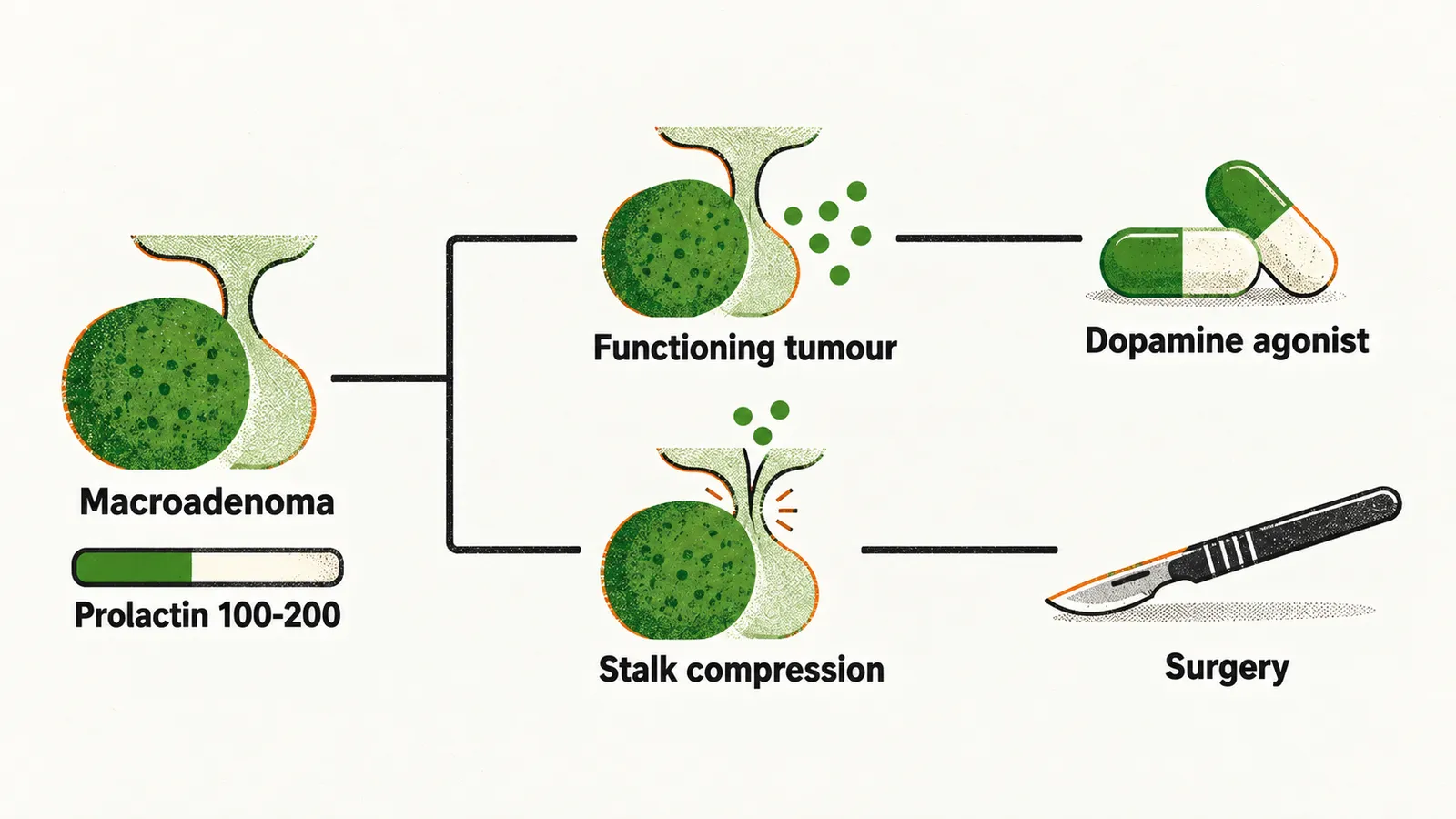
If a patient has a macroadenoma but the serum level of prolactin is less than 200 ng/ml it can be:
- if the laboratory measurement of prolactin is reliable, this is a pseudo-prolactinoma
- if the laboratory measurement of prolactin is not reliable, it could be a macroadenoma
- if more than 50% of the prolactinoma is a cystic sellar mass, then usually in these patients the size of the prolactinoma is not correlated with the level of serum prolactin, which is low
The reliability of the laboratory measurement therefore decides how a low level is read, and it can fail in several ways.
Stress and serial sampling
Similar to GH (growth hormone), prolactin secretion is also affected by stress; therefore a laboratory-based test for the measurement of prolactin is better done in serial form to prevent the effect of stress-induced secretion of prolactin in response to venipuncture.
Laboratory pitfall: the high-dose hook effect
A laboratory result for prolactin can also be incorrect because of the assay itself. In some laboratory techniques it is possible that a very high concentration of prolactin in the serum saturates both the signal and the capture antibodies, so the result from the signal antibodies is not correct, which leads to under-estimation of the serum prolactin level. This effect is called the high-dose hook effect.
In cases with suspicion of the high-dose hook effect the test can be repeated with dilution of the patient’s serum at 1:100.
Laboratory pitfall: macroprolactinemia
Other than the high-dose hook effect as a laboratory pitfall in the diagnosis of hyperprolactinemia, there is another pitfall, in which patients do not have any clinical signs and symptoms of hyperprolactinemia, but the laboratory results show significant hyperprolactinemia. Studies of these patients show that the hyperprolactinemia is caused by a big-big prolactin aggregate, since the biological activity of prolactin is exerted by the monomeric form of prolactin and not by the aggregate molecules. These patients are called macroprolactinemia.
Big-big prolactin aggregates are molecules which can be found in the serum due to the formation of a complex between IgG (immunoglobulin G), or IgA, and the circulating prolactin, which makes prolactin aggregates that have a high molecular weight in comparison to the monomeric form, which is the active form of the hormone. These antibodies are autoantibodies against different epitopes of prolactin, which can differ in different individuals, with different association rates with the antigen, which is prolactin. Therefore, the condition of macroprolactinemia is defined as the presence of big-big prolactin in the serum which predominates over other forms by 30-60%.
So, in a patient without any clinical signs and symptoms of hyperprolactinemia but with laboratory results showing macroprolactinemia, a precipitation test of the serum with PEG (polyethylene glycol) has to be performed; its positivity shows that the high level of prolactin is made by the big-big prolactin aggregate, which is not biologically active, and that there is no adenoma in these patients.
PCOS as a differential diagnosis
A very important differential diagnosis to consider in women with hyperprolactinemia is PCOS (polycystic ovary syndrome). In about 15% of women with PCOS there is hyperprolactinemia on the laboratory result, but much of it reflects macroprolactinemia, the biologically inactive big-big aggregate, rather than a true excess of the active hormone. A prolactinoma explains a minority of these elevations, so it is looked for when a true hyperprolactinemia persists after the artefact has been excluded.