Confirming hypopituitarism requires testing the pituitary axes both at rest and after stimulation. The approach is to check both the basal and the stimulated, or dynamic, level of each hormone.
Baseline testing
To evaluate the basal serum concentration of the anterior pituitary hormones and the hormones formed by their target glands, a blood sample is taken in an unstressed condition between 7 and 9 am, the window in which the serum levels of cortisol and testosterone are highest.
| Axis/system | Tests |
|---|---|
| Adrenocortical | Morning serum cortisol, ACTH |
| Thyroid | Free T4, TSH |
| Gonadal - men | Testosterone (09.00 AM), SHBG, LH, FSH |
| Gonadal - women | Estradiol, LH, FSH, progesterone (day 21 if menstruating) |
| Prolactin | Prolactin level |
| Growth | Insulin-like growth factor-1, growth hormone |
| Fluid balance | Paired plasma and urine osmolality |
The adrenal axis
In central hypoadrenalism, or secondary hypoadrenalism, ACTH can be low or low-normal, so a low or low-normal ACTH does not exclude the diagnosis. ACTH remains the gold-standard biomarker for distinguishing primary from secondary hypoadrenalism, because in primary disease the ACTH level is inappropriately high.
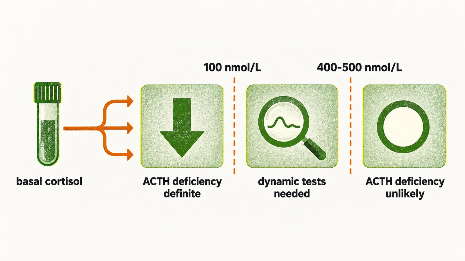
When ACTH deficiency is suspected, the basal cortisol at 9 am determines the next step:
- below 100 nmol/L (3.625 µg/dL) → ACTH deficiency is definite
- above 400-500 nmol/L (14.5-18.1 µg/dL) → ACTH deficiency is unlikely
- between 100 nmol/L (3.625 µg/dL) and 400-500 nmol/L (14.5-18.1 µg/dL) → dynamic tests are needed to confirm it
When the basal cortisol falls in this grey zone, the insulin tolerance test (ITT) can be used; induced hypoglycaemia stimulates the axis, and the result is interpretable only if the induced glucose fell below 40 mg/dL. When the ITT is contraindicated in a patient with suspected secondary hypoadrenalism, the ACTH stimulation test is performed instead. In normal subjects the response to the ACTH stimulation test raises the blood cortisol above 500 nmol/L (18 µg/dL), the traditional threshold for a normal response.
Growth hormone testing
The ITT is also the test of choice for growth hormone. Several tests exist for measuring the dynamic changes of the different pituitary hormones, but the ITT is still preferred, because it measures the dynamic changes of ACTH and GH at the same time. Insulin-induced hypoglycaemia is a potent stimulator of pituitary hormone release, which is why the ITT can be used to measure the reserve of both GH and ACTH.
When the ITT is contraindicated in a patient with suspected growth hormone deficiency (GHD), the recommended test is the combination of arginine and growth hormone-releasing hormone (GHRH), and its result is judged against the patient’s body mass index (BMI):
- BMI below 25 → GH below 11 microgram/L
- BMI between 25 and 30 → GH below 8 microgram/L
- BMI above 30 → GH below 4 microgram/L
For the diagnosis of GHD, IGF-1 as a biomarker is highly specific but not sensitive: a low IGF-1 level is enough to confirm GHD, but a normal or low-normal IGF-1 is quite common in patients with GHD, so it cannot rule the diagnosis out. The overlap is probably explained by the fall in IGF-1 during physiological aging, which brings the age-adjusted reference values and the pathological decrease of GHD close together.
The thyroid axis
The thyroid axis is assessed with basal measurements, and the difficulty lies in interpreting them: the two measurements are TSH and free thyroxine (free T4, abbreviated FT4).
TSH can be measured directly because of its relatively long half-life. Suspected secondary hypothyroidism, however, cannot be approached in the same way as primary disease. In primary disease the laboratory method starts with the TSH: if it is high or low, FT4 follows, and the combination guides the diagnosis of primary hyper- or hypothyroidism. In central hypothyroidism the great majority of patients have a TSH that is normal or even slightly increased, and the reason is the formation of biologically inactive TSH by the anterior pituitary: enough TSH is present, but it is not biologically active. The method for diagnosing central hypothyroidism is therefore the simultaneous measurement of TSH and FT4, rather than checking TSH first and following with FT4 only when it is abnormal. Based on the guidelines, mild central hypothyroidism is diagnosed when there is a borderline FT4 with an inappropriately low or low-normal TSH, together with some other supportive evidence in the patient.