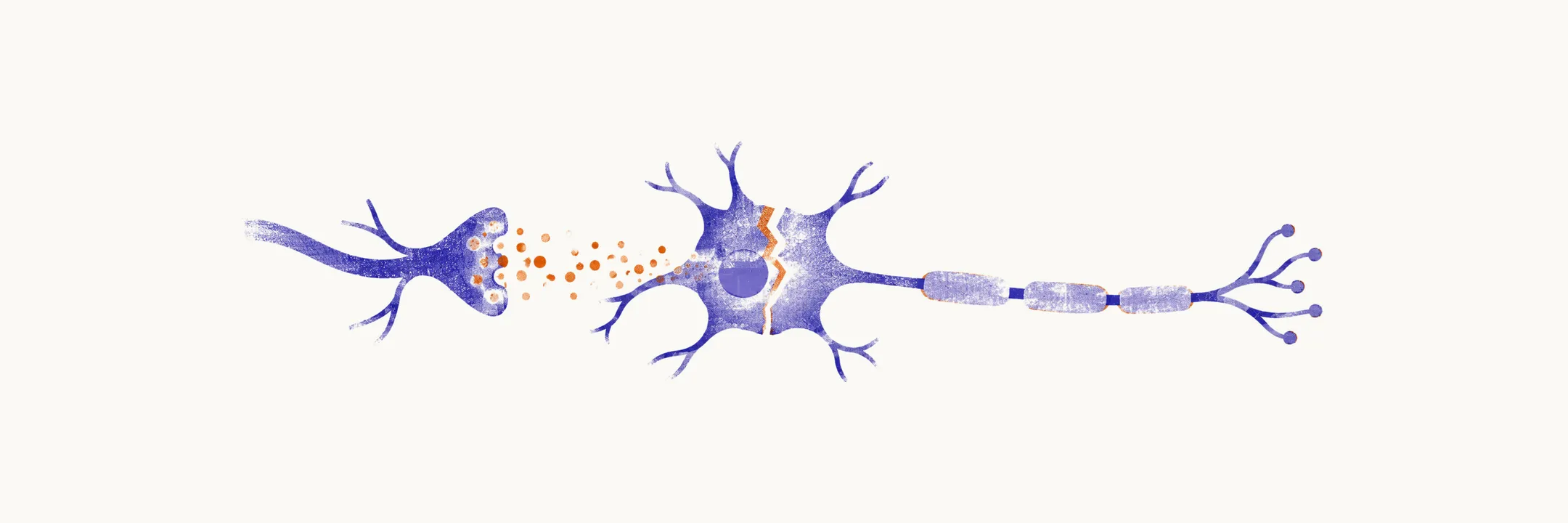
ALS destroys upper motor neurons (UMN), which begin in the motor cortex, and lower motor neurons (LMN), which lie in the brainstem and spinal cord and connect to muscle. Sensory, autonomic, and most cognitive systems stay largely intact. Why the motor system fails first remains incompletely understood, and the mechanisms below are best read as partial explanations.
Why motor neurons are vulnerable
Motor neurons are selectively damaged, and three hypotheses try to explain why. None is proven, and they likely combine.
- Axonal length. Motor neurons are among the largest cells in the body. Axons supplying leg muscles can exceed a metre. That length imposes transport and energy demands that may leave the cell with little reserve.
- Cortical origin (“dying forward”). Some evidence places the start in the motor cortex, with disease spreading anterogradely to spinal motor neurons. This model remains debated.
- Combined risk. Genes, metabolism, and environmental exposures appear to interact in susceptible cells. No single factor explains sporadic disease.
Glutamate and oxidative injury
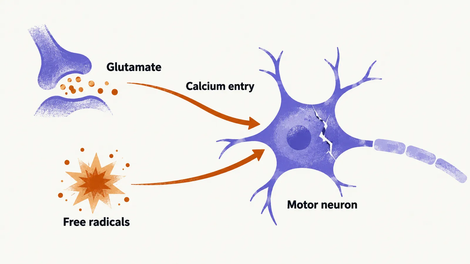
One suspected route of injury is excess synaptic glutamate, the main excitatory neurotransmitter. It overstimulates motor neurons, and the resulting calcium entry kills the cell. This sequence is called excitotoxicity. Riluzole is believed to reduce this damage by decreasing glutamate release, though its exact action remains uncertain.
A second route is oxidative stress. Motor neurons also face damage from free radicals, which are reactive molecules that injure cell components. Edaravone is thought to neutralise these radicals. Its clinical benefit is small and limited to selected patients.
TDP-43 and the link with frontotemporal dementia
Neurons in ALS also accumulate abnormal protein. In 2006, TDP-43 was identified as the deposited protein in most ALS and in the common behavioural forms of frontotemporal dementia (FTD), a dementia that mainly changes behaviour, personality, or language. The two diseases are now seen as different expressions of one TDP-43 proteinopathy, meaning a disease defined by a deposited protein. About 97% of motor neuron disease shows this molecular pathology.
In cohorts, roughly half of ALS patients develop cognitive or behavioural change. About 10% develop full FTD. A comparable share of FTD patients develops motor signs over time. The C9orf72 repeat expansion is the most common genetic cause of both.
Genes: C9orf72 and SOD1
Genetics gives a second link between ALS and FTD. In familial ALS, meaning ALS that runs in families, C9orf72 hexanucleotide expansions cause about 25–40% of cases. SOD1 mutations cause another 12–20% of familial cases, roughly 2% of all ALS.
Genetic testing is offered when family history, young onset, or FTD features suggest inherited disease. A confirmed SOD1 mutation is required before tofersen treatment. Prevention trials in carriers without symptoms, such as the ATLAS study, are ongoing and not established care.
Whatever the cause, ALS does not look the same in every patient: which motor neurons fail first shapes how the disease presents and how fast it progresses.