Cushing’s syndrome is confirmed biochemically before its cause is sought. The reason for that order is practical: the clinical features are non-specific and the causes are diverse, so the diagnosis is built first from tests that show the cortisol excess itself, and only then from tests and imaging that separate the ACTH-dependent from the ACTH-independent causes.
When to suspect Cushing’s syndrome
Because single clinical features are non-specific, Cushing’s syndrome has to be suspected before the diagnostic approach begins. The situations that should raise that suspicion are:
- hypertension in young adults, or resistant hypertension
- poorly controlled diabetes despite appropriate therapy — about 4% of the patients with type 2 diabetes mellitus are suffering from Cushing’s syndrome
- fragility fractures in eugonadal subjects (those with normal gonadal function)
- decreasing growth velocity or weight gain in children
- adrenal incidentalomas (adrenal masses discovered by chance on imaging done for another reason)
- signs of hypercortisolism — striae, muscle weakness and others
- use of exogenous glucocorticoids
The two challenges and the role of pre-test probability
The diagnosis presents two main challenges: distinguishing a pathological increase of glucocorticoids from the physiological causes of a high level of cortisol production, and then determining the etiological cause of the pathological excess.
The diagnosis of Cushing’s syndrome is considered established if the results of several tests are consistently suggestive of Cushing’s syndrome. How many tests are needed depends on how likely the diagnosis was before testing: when the pre-test probability is mild to high, 2-3 screening tests should be used, while in a patient with a low pre-test possibility a single negative test is enough to exclude the syndrome.
First-line screening tests
The screening tests fall into three groups, each of which exploits a different normal property of the hypothalamic-pituitary-adrenal axis that is lost in Cushing’s syndrome.
Urinary free cortisol
The most commonly used screening test is the urinary concentration of cortisol, measured in 2-3 different 24-hour collections of urine for free cortisol, or UFC, and it is the first line of screening. Its sensitivity for hypercortisolemia is about 95% in patients with Cushing’s disease and about 88% in ectopic forms and adrenal masses.
Two practical points govern its interpretation. First, the collection must be complete: creatinine excretion is measured to check this, and it should be about 1 g/day in a patient of about 70 kg. Second, the ratio between cortisol and creatinine is more informative than cortisol alone, and a cortisol-to-creatinine ratio of more than 25 nmol/mmol is suggestive of Cushing’s syndrome.
The advantage of the UFC test is that it reflects only the free cortisol in the blood, not the cortisol bound to transcortin, also called corticosteroid-binding globulin (CBG). In patients whose CBG level is altered, the UFC test is therefore more specific for hypercortisolism than tests performed on serum.
Loss of the negative feedback on ACTH secretion
Physiologically, cortisol feeds back negatively on CRH (corticotropin-releasing hormone) and vasopressin, the hormones that stimulate ACTH release, and in patients with Cushing’s syndrome this negative feedback is resisted.
Dexamethasone can be used to test that feedback, and it has been used in two ways. The low-dose dexamethasone suppression test (LDDST) gives a low dose of dexamethasone and then measures glucocorticoid in the urine. The overnight dexamethasone suppression test (ONDST) gives 1 mg of dexamethasone during the night, at 11 pm, and measures the plasma cortisol at 9 am. Because it is easier, the ONDST is the one used today, and it is the most sensitive test among all of these, with a sensitivity of 98-99% and a cut-off of 50 nmol/L.
Loss of the circadian rhythm
Cortisol secretion normally follows a circadian rhythm, with a peak around 7-8 in the morning and a low level during the night. In Cushing’s syndrome this rhythm is disrupted: in some patients the morning peak remains, but in most the midnight nadir is lost and the cortisol level stays high at midnight. This is why any screening test performed at midnight has a high diagnostic value.
Midnight salivary cortisol is the convenient way to sample that time point. It is very easy to perform and requires no invasive step, and studies show a sensitivity of 92% and a specificity of 96%.
Pitfalls in interpreting the tests
Several factors can distort these results when a diagnosis rests on them:
- An incomplete 24-hour urine collection, containing more or less than the full day, gives a misleading UFC.
- A CYP3A4-inducing drug, such as rifampicin or an antiepileptic, increases the metabolism of dexamethasone and so weakens the dexamethasone test.
- An oral contraceptive increases the CBG level and with it the total cortisol, which can mask the effect of the dexamethasone test; if so, the test should be repeated 4-6 weeks after the withdrawal of estrogen.
Pseudo-Cushing’s syndrome
A further source of confusion is pseudo-Cushing’s syndrome, the state in which a patient has both the clinical features of Cushing’s syndrome and some biochemical evidence of hypercortisolemia, but recovers once the predisposing factor is resolved. It occurs mostly in patients with depression, alcohol abuse, pregnancy, morbid obesity, or poorly controlled diabetes mellitus.
Second-line screening tests
When the first-line tests and their pitfalls leave the diagnosis unclear, two second-line screening tests can be used: one that samples the midnight nadir directly and one that tests the pituitary’s response to desmopressin.
Midnight serum cortisol was, before the introduction of salivary cortisol, the only reliable test for the midnight nadir. It is hard for the patient, who has to stay in the inpatient clinic for at least 48 hours, and the sample should be taken 5-10 minutes after waking the patient during the night. A plasma level of less than 50 nmol/L excludes Cushing’s syndrome. False positive results can occur in patients who are critically ill, have acute infections, or have heart failure. Late-night salivary cortisol is thus an outpatient test, while midnight serum cortisol is an inpatient test.
The desmopressin test is the other second-line option. Desmopressin leads to a rise in ACTH and cortisol because the corticotroph cells of the anterior pituitary, and corticotroph adenomas, carry V3 receptors that desmopressin can activate.
Separating ACTH-dependent from ACTH-independent disease
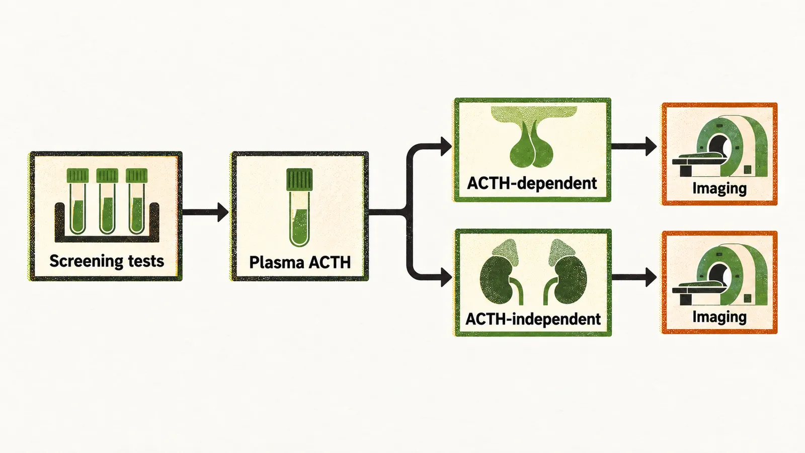
Once the tests above have confirmed hypercortisolemia, the next step is the differential diagnosis between ACTH-dependent and ACTH-independent disease, and it begins with a measurement of the level of ACTH in the plasma: if it is inappropriately normal or increased, the disease is ACTH-dependent.
The sample for plasma ACTH should be taken at about 8-9 am, and it is suggested to test twice, because some patients show an occasional low plasma ACTH of less than 10 ng/L. The interpretation follows the level:
- a plasma ACTH of less than 10 ng/L suggests ACTH-independent Cushing’s syndrome
- a plasma ACTH consistently more than 20-30 ng/L suggests either Cushing’s disease or ectopic Cushing’s syndrome
- an intermediate level, between 10 and 20-30 ng/L, should be studied further with the CRH or desmopressin test.
Imaging
Imaging should be performed only after these findings, because the ACTH result narrows it to specific organs. In ACTH-independent disease the imaging is of the adrenal gland, and an un-enhanced CT scan (one without contrast) is used. In ACTH-dependent disease the imaging is MRI of the pituitary gland, and adrenal imaging is not indicated. The limitation to keep in mind is that in about 40% of cases the pituitary adenoma is missed on MRI because of its size.
High-dose dexamethasone, CRH and inferior petrosal sinus sampling
In ACTH-dependent Cushing’s syndrome a biochemical test is always needed after the imaging to distinguish ectopic production from a pituitary adenoma, because a pituitary adenoma retains some response to dexamethasone for negative feedback, while ectopic disease mostly does not.
Mostly in clinical cases the patient is first tested with the high-dose dexamethasone suppression test (HDDST), and then the high-dose dexamethasone test is repeated; if the cortisol is resistant to suppression on the second test, ectopic ACTH production is probable. A positive response is defined as suppression of the plasma cortisol level to less than 50% of the basal level.
If the HDDST and the CRH test give incompatible results, the inferior petrosal sinus sampling (IPSS) can be used. It is based on the injection of CRH and the comparison of the petrosal/peripheral ACTH ratio at baseline and 5 minutes after injection. It has very high sensitivity and specificity for Cushing’s disease when the petrosal/peripheral ACTH ratio is greater than 2 at baseline and greater than 3 five minutes after injection. IPSS is also used to confirm Cushing’s disease when MRI is not sensitive enough to detect an adenoma of less than 10 mm.
The ectopic sources
When the differential points toward ectopic ACTH-dependent Cushing’s syndrome, a CT scan of the chest and abdomen is performed to locate the tumour. If no lesion is found there, MRI of the chest or abdomen can be used, which may show a carcinoid lesion better. The most common sources of ectopic ACTH release are bronchial neuroendocrine tumours, which account for 25% of ectopic ACTH-dependent Cushing’s syndrome, and small cell lung carcinoma, which accounts for 19%.
ACTH-dependent Cushing’s syndrome and ectopic carcinoma commonly have hyperandrogenism alongside the high glucocorticoid level, whereas in ACTH-independent Cushing’s syndrome there is mostly only the high level of glucocorticoid.