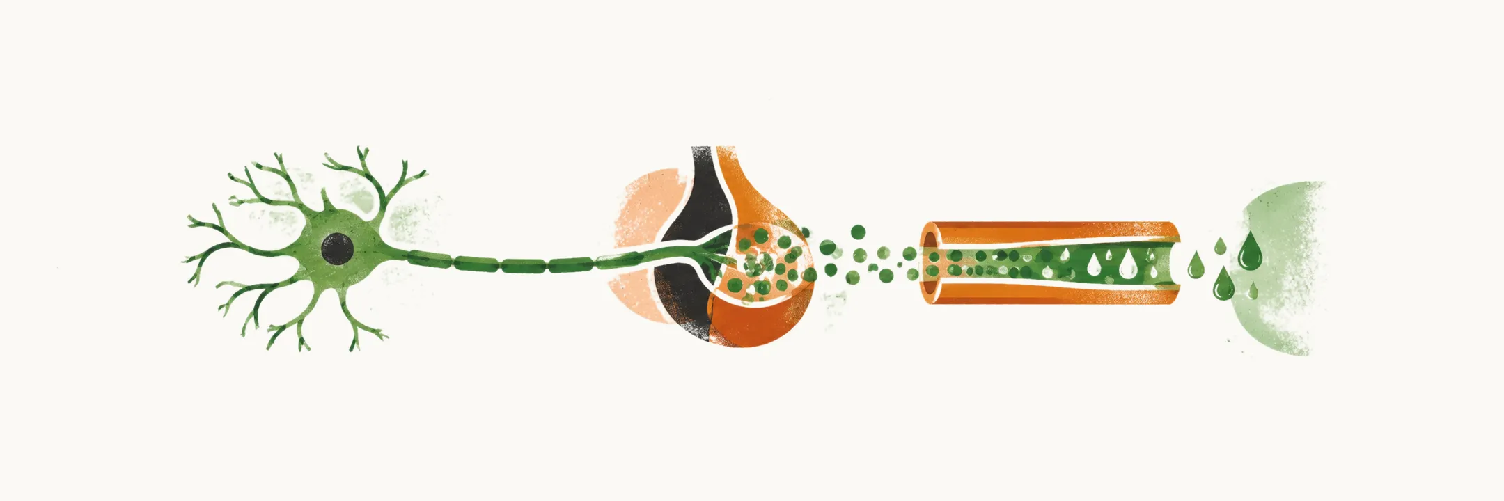
Antidiuretic hormone (ADH), also called arginine vasopressin (AVP), is the hormone that lets the kidney save water. Its release is set by how concentrated the blood is, and its target is the collecting duct of the kidney, where it opens water channels so that water returns to the body instead of being lost in the urine.
Where ADH is made and released
ADH is a small peptide made by magnocellular neurons, large nerve cells whose cell bodies sit in two nuclei of the hypothalamus: the supraoptic nucleus and the paraventricular nucleus. These neurons send their axons down the pituitary stalk and end in the posterior pituitary, where the hormone is stored in nerve terminals and released into the bloodstream when the neurons fire. Because the cell bodies lie in the hypothalamus while the release site is the posterior pituitary, damage at any point along this path — the hypothalamus, the stalk, or the gland — can reduce ADH release. This shared pathway is why the resulting disease is called hypothalamic, or central, diabetes insipidus.
How release is controlled
The magnocellular neurons carry osmoreceptors: they sense the osmolality of the plasma, that is, its concentration of dissolved particles, measured in milliosmoles per kilogram of water (mosmol/kg). When plasma osmolality falls below a threshold of about 280-285 mosmol/kg, ADH release is suppressed, so the kidney keeps producing dilute urine; when osmolality rises above it, ADH release increases and the urine is concentrated. Thirst is regulated in parallel and at a similar threshold, so a healthy person both drinks and retains water to defend plasma osmolality. Blood volume and pressure form a second, less sensitive control: a large loss of blood volume stimulates ADH release powerfully even when osmolality is normal, because retaining water is part of defending the circulation.
How ADH concentrates urine
ADH reaches the kidney and binds the V2 receptor on the principal cells of the collecting duct, the cells that control how much water is reabsorbed from the forming urine. The signal inserts aquaporin-2 water channels into the apical membrane of these cells, the membrane facing the urine, so water leaves the forming urine and returns to the body, leaving a small volume of concentrated urine. When ADH falls, the channels are withdrawn from the membrane and the urine stays dilute.
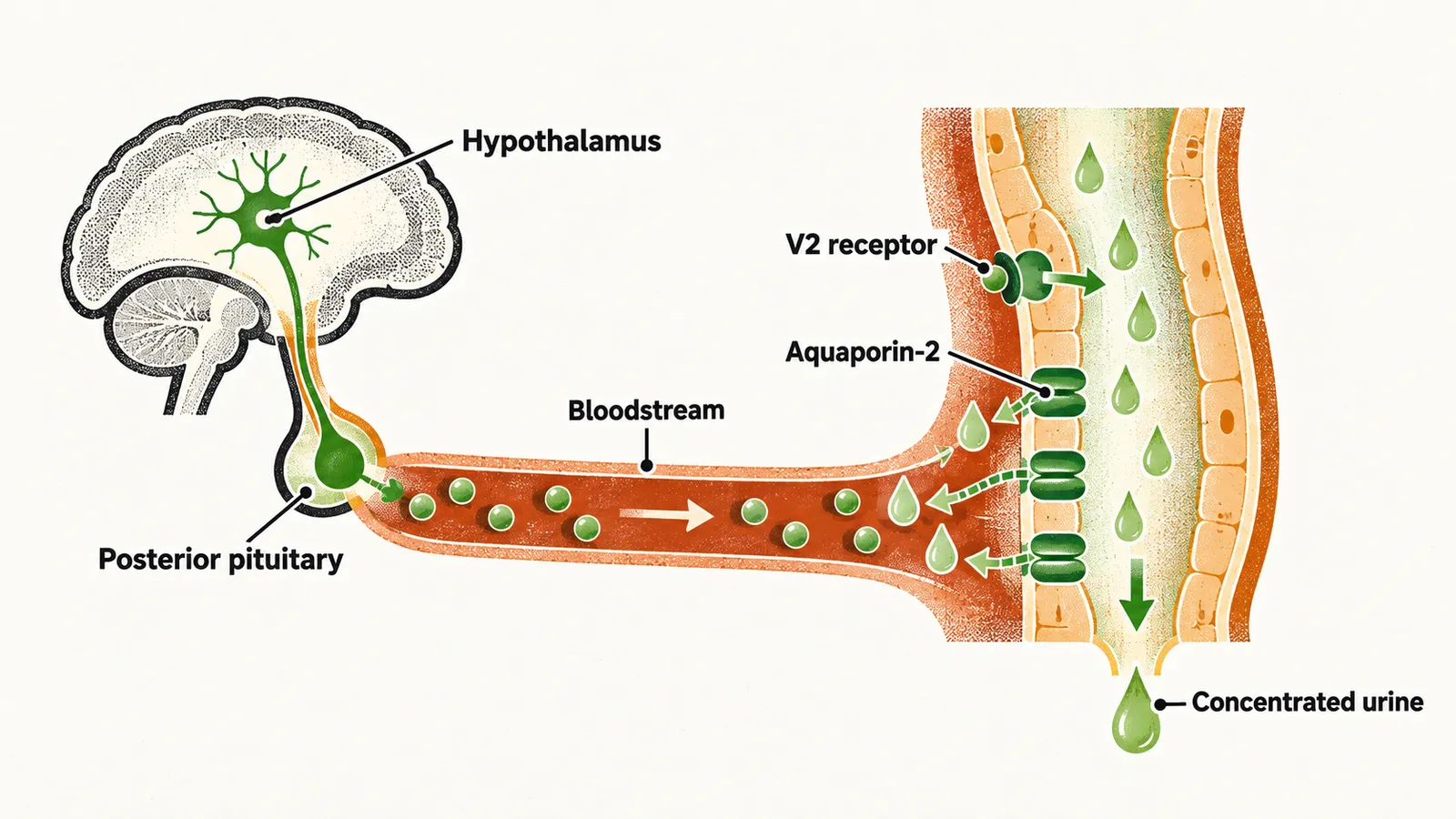
Urine concentration therefore depends on two things at once: enough ADH reaching the kidney, and a kidney able to respond to it. Lose the first and the result is central diabetes insipidus; lose the second and the result is nephrogenic diabetes insipidus.