Hypothalamic diabetes insipidus is the form of diabetes insipidus that results from too little secretion of antidiuretic hormone (ADH, also called arginine vasopressin or AVP), the hormone that lets the kidney concentrate urine. It is also called central diabetes insipidus.
Mechanism
In hypothalamic diabetes insipidus, or HDI, the secretion of ADH is not enough to concentrate the urine, so hypotonic polyuria develops and the patient passes large volumes of dilute urine, with nocturia (waking at night to pass urine) as well. The magnocellular neurons that produce AVP are numerous, so the disease appears only after most of them are lost: more than 80% of the hypothalamic AVP neurons must be destroyed before diabetes insipidus becomes manifest. Because the thirst mechanism remains intact in most cases, patients drink enough to replace the water they lose and the plasma osmolality (the concentration of dissolved particles in the plasma) stays normal, so the disorder is usually found through the polyuria rather than through a raised plasma sodium.
Prevalence and the surgical setting
HDI is generally very rare, with a prevalence on the order of 4 in every 100,000 of the population, but it is fairly common among patients who undergo neurosurgery for pituitary tumours. Post-operative HDI occurs in 18-30% of these operations. In most cases the natural history is spontaneous resolution over 2-5 days after surgery, while in about 1-8% the HDI remains permanent.
A plasma sodium of more than 145 mmol/l after neurosurgery, which carries a significant risk of HDI, shows a specificity of more than 98% for the diagnosis of post-operative HDI.
The triple-phase course
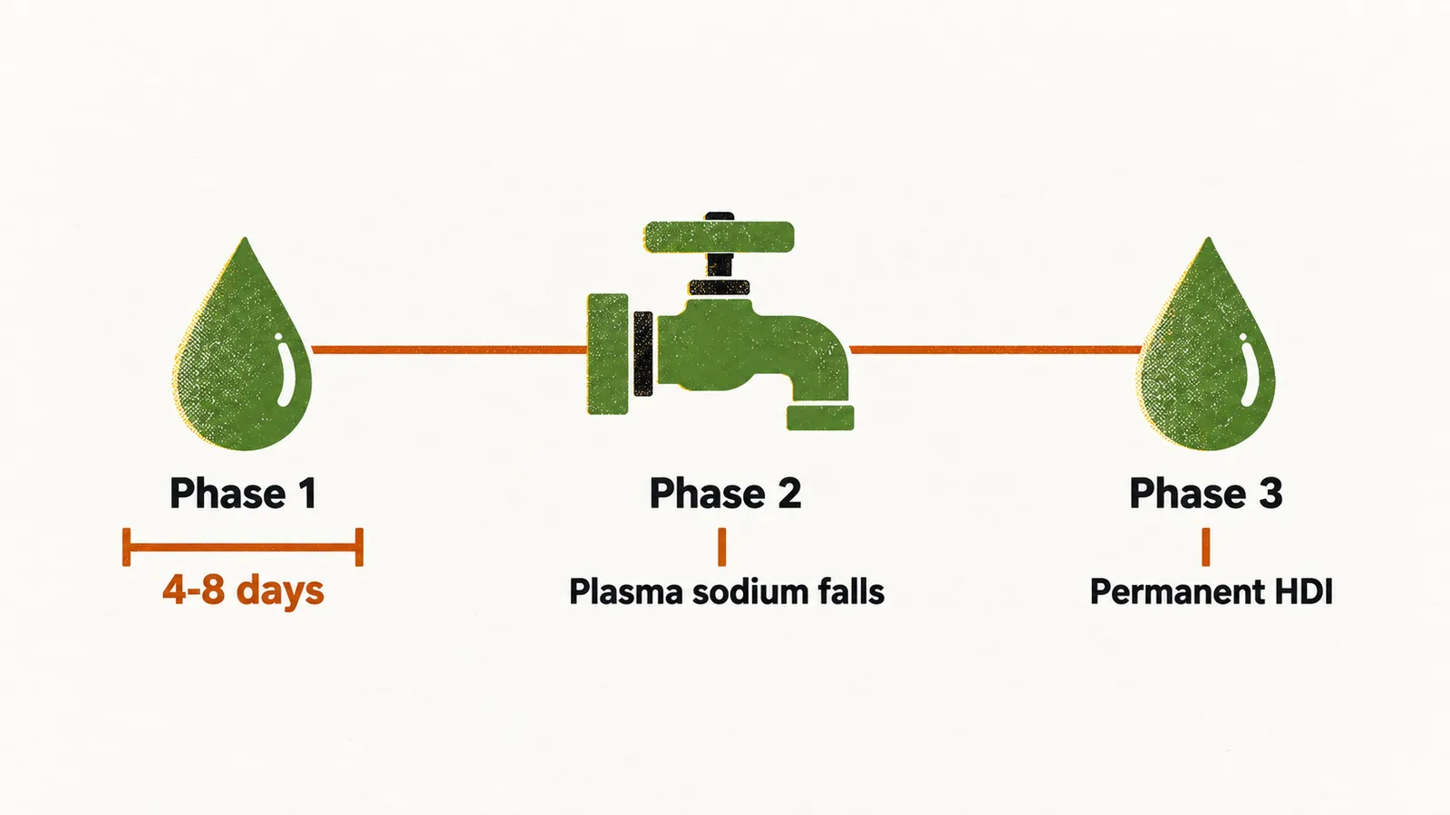
When post-operative HDI is permanent, it often follows a stereotyped sequence called triple-phase HDI:
- Phase 1: polyuria and hypotonic urine for 4-8 days after surgery, with trace amounts of AVP in plasma.
- Phase 2: the damaged magnocellular neurons release the AVP they had stored, without the normal feedback control, so the kidney retains water and the plasma sodium can fall — a picture of inappropriate antidiuresis.
- Phase 3: permanent HDI.
Other causes
Surgery is the commonest setting, but not the only one. Pituitary masses rarely cause diabetes insipidus by themselves. When they do, the most common pathologies are craniopharyngiomas (tumours of the sellar region, around the pituitary), hypophysitis (inflammation of the pituitary) and granulomas.
Autoimmune diabetes insipidus
Idiopathic diabetes insipidus, meaning a case with no identified cause, used to be diagnosed more often, but after the introduction of more sophisticated imaging techniques and greater awareness of the autoimmune conditions that can affect the CNS — especially the hypothalamus and the pituitary gland — it is now known that most of the idiopathic cases were autoimmune. Autoimmune diabetes insipidus is characterised by:
- young age of onset;
- circulating autoantibodies against the AVP-secreting cells;
- a thickened pituitary stalk on T1-weighted MRI.
Patients with autoimmune diabetes insipidus more commonly have autoimmune conditions of other organs as well; in about 30% of the cases there is also autoimmune disease of the thyroid gland.
Traumatic brain injury
In 20% of cases of traumatic brain injury there is diabetes insipidus, and in nearly all of them the condition is transient. When it is permanent, the course is a triple-phase response similar to the permanent diabetes insipidus that follows surgery.