
Appetite is set in the brain, which weighs signals of hunger and satiety coming from the body. Obesity is therefore not just a metabolic disorder: its progression and pathogenesis may have a neuropsychological basis.
Homeostatic and non-homeostatic control of eating
Eating is controlled by anorexigenic, or satiety, signals and orexigenic, or hunger, signals that reach the brain and are interpreted there as the desire to eat. The signals are of several kinds: blood-borne, such as the blood glucose level; hormonal, such as leptin and insulin; and neurotransmitter, such as dopamine.
The brain regions that control hunger fall into two groups. The older circuits — the hypothalamus and midbrain — are homeostatic; they are found in all mammals and are preserved through evolution. The newer circuits — cortical regions sitting above the older ones — are non-homeostatic; they control the older circuits and give feedback to them, and they are found in humans.
The arcuate nucleus and its signals
The main region that responds to anorexigenic and orexigenic signals lies in the hypothalamus, in the arcuate nucleus. It receives three signals in particular.
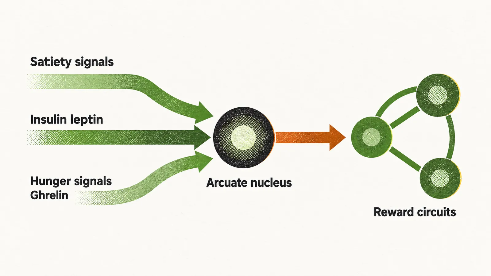
Insulin is physiologically a satiety signal, but in obese patients the CNS (central nervous system) is resistant to it because of chronic hyperinsulinaemia, a persistently high insulin level, so the suppressive effect of insulin on hunger is lost. Leptin is also a satiety signal, and it fails in the same way: the chronically high leptin level makes the CNS resistant to leptin, so its suppression of appetite is lost too. Ghrelin acts in the opposite direction. Physiologically it is an orexigenic signal that induces hunger, and eating normally stops its release from the oxyntic cells of the stomach; in obese patients, however, that fall in release after food intake does not occur.
Reward circuits
Besides the arcuate nucleus, the other important homeostatic region of the brain in appetite control is the limbic system, especially the amygdala and hippocampus. These regions are under the control of dopaminergic neurons that form a reward-based system. In obese patients one can speak of addiction to food, or at least to some particular foods, which induce the release of dopamine from these regions and manipulate hunger and appetite through reward. The same hormonal factors that act on the arcuate nucleus act here as well: leptin and insulin inhibit the dopaminergic neurons, while ghrelin stimulates dopamine release.
GLP-1
GLP-1 (glucagon-like peptide-1) is a further hormonal factor in the control of appetite, alongside insulin, leptin and ghrelin. It is released by the L-cells of the intestine and by some cells in the brain in response to food intake, and it reduces appetite, delays stomach emptying and increases intestinal secretion, among other effects. The GLP-1 released in the brain comes from neurons of the nucleus tractus solitarius, or NTS; it acts on the ventral tegmental area (VTA) to reduce high-fat diet intake, and on the hypothalamus and the nucleus accumbens.
Cortical control
Above the older neuronal regions, newer regions in the cortex also affect appetite. One of the most important is the orbitofrontal cortex, which suppresses food intake; in obese patients this region is not able to decrease appetite strongly enough.
Failure of these braking signals is one route by which fat accumulates and persists; what that excess fat then does to the body is the next question.