
A suspected pituitary tumor is investigated in four ways: magnetic resonance imaging (MRI) shows the mass and its surroundings, ophthalmologic evaluation tests the vision it threatens, laboratory investigation looks for hormone excess or loss, and histologic investigation examines the tissue when the other results do not settle the diagnosis.
Magnetic resonance imaging
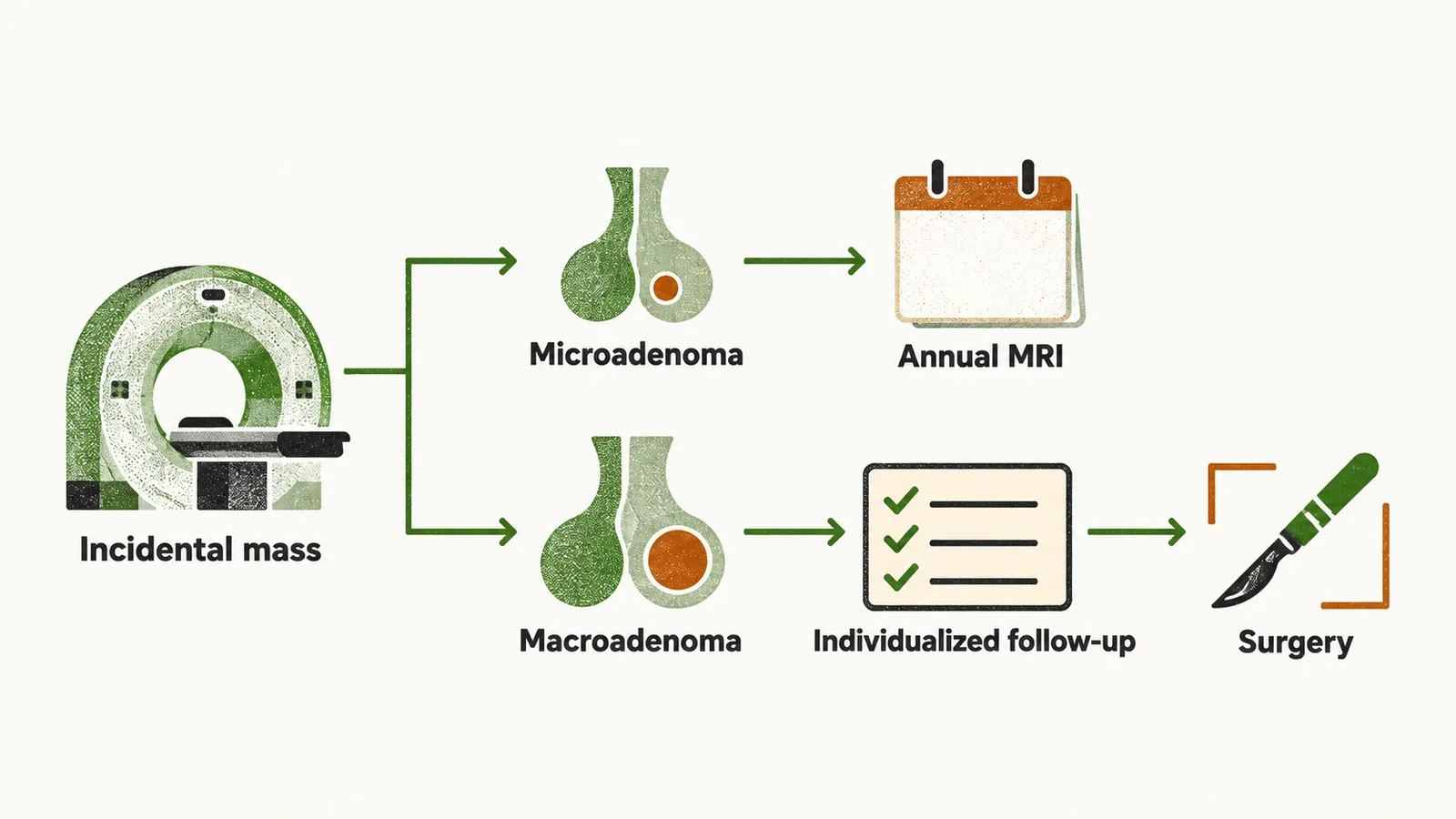
Sellar masses are mostly incidental findings on MRI, and most are pituitary adenomas, which are then called incidentalomas, meaning masses discovered by chance in a scan done for another reason. What to do with one depends on its size and on whether it causes any effect:
- In the absence of hypersecretion, an incidental microadenoma is simply followed with an annual MRI without any intervention in the patient.
- An incidental macroadenoma without mass effect or visual compromise does not automatically need surgery. It is managed with individualized follow-up, with hormonal, imaging and visual-field assessment, and surgery is offered when the tumor compresses the optic chiasm, causes other neurological compromise, or turns out to be a hypersecreting tumor.
For the image itself, sagittal and coronal T1-weighted MRI before and after administration of gadolinium, a contrast agent, provides a precise picture of the hypothalamus, the pituitary stalk, the pituitary tissue, the cavernous sinus, the sphenoid sinus and the optic chiasm.
Normal anterior pituitary gland soft tissue on MRI resembles heterogeneous tissue. The density of an adenoma is usually different from that of the surrounding tissue. CT scan is reserved for investigation of the level of bone erosion and also calcification of the mass.
Ophthalmologic evaluation
Because the optic chiasm lies next to the gland, the eye examination can show the effect of a mass that imaging only outlines. The classical ophthalmological finding in pituitary tumors is bitemporal hemianopia, which is partial blindness of the outer vision. It is more pronounced for superior vision. This happens because the nasal ganglion cell fibers of the retina are more vulnerable to compression of the ventral optic chiasm.
Invasion of the tumor into the cavernous sinus can produce diplopia due to invasion of the ocular motor nerves that are located in the cavernous sinus.
Laboratory investigation
In the functional pituitary adenomas, which are Cushing’s disease, prolactinoma, and acromegaly, the clinical manifestation should guide the laboratory investigation.
Usually, when a pituitary tumor is suspected on MRI, several laboratory tests are performed:
- prolactin (PRL)
- IGF-1 (insulin-like growth factor 1)
- 24-hour urinary free cortisol
- FSH and LH
- thyroid function tests
Histologic investigation
Histological staining and molecular studies of the specimen help make the diagnosis in the cases that have equivocal hormone results and also in the case of non-functioning tumors.