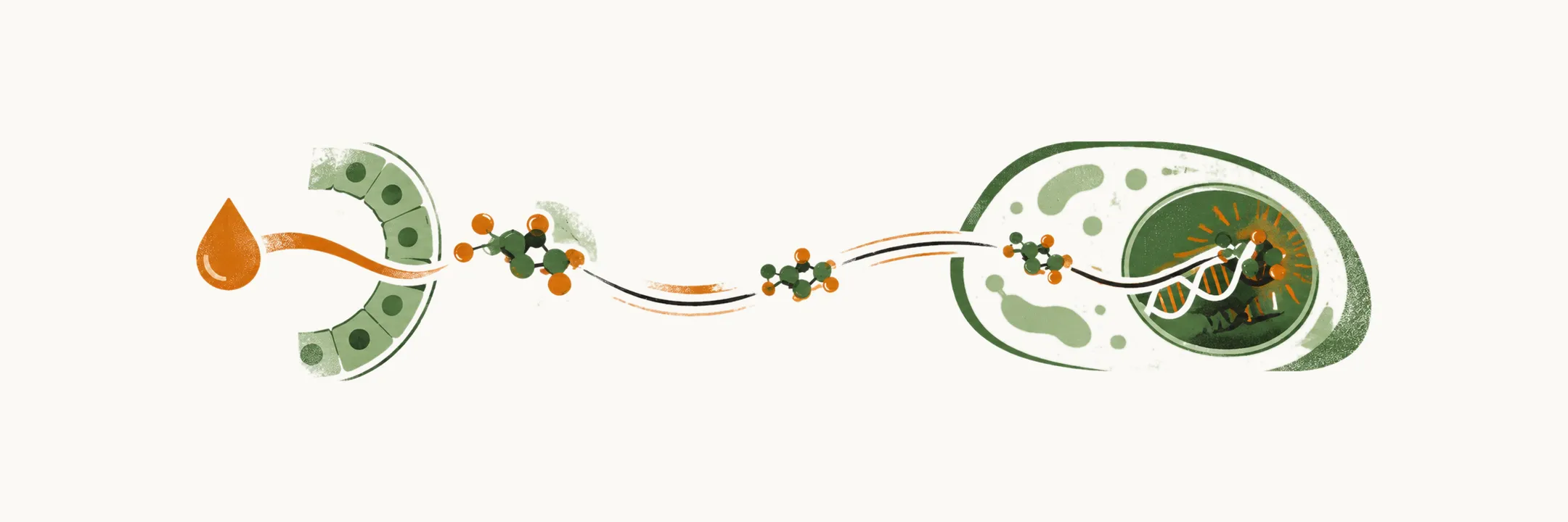
The thyroid gland builds two hormones, thyroxine (T4) and triiodothyronine (T3). Producing them takes iodine, a set of enzymes and the protein scaffold thyroglobulin; once they are released, they are carried in the blood and converted to their active form at the tissues.
Thyroid hormone metabolism
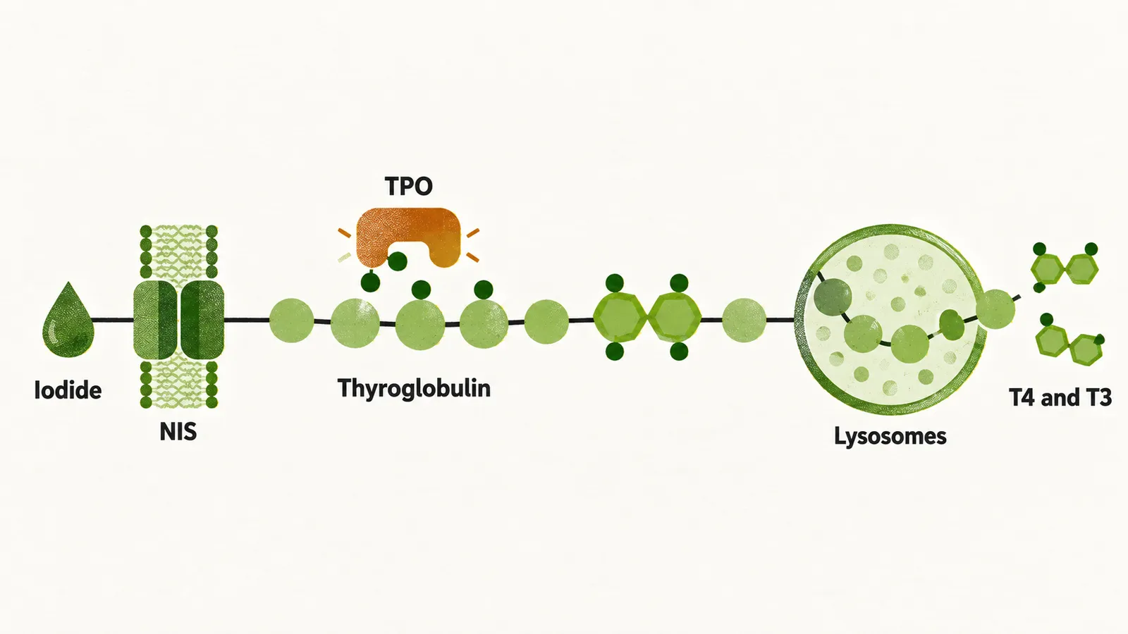
The first step in the production of thyroid hormone is the absorption of iodine by the follicular cells of the thyroid gland. This absorption is done in a very efficient manner by the sodium–iodide symporter (NIS), a membrane transporter that carries iodide into the cell and is located on the basolateral side of the follicular cells. The expression of NIS depends on the level of iodine in the blood: the lower the amount of iodine, the higher the expression of NIS, and the reverse. That dependence is the basis both of thyroid scanning for diagnosis and of the use of radioactive iodine for treatment, because the gland takes up the tracer or the therapy in proportion to how much iodine it is seeking. Besides NIS, another transporter, pendrin, is located on the apical surface of the follicular cells, the side facing the lumen.
Several regions in the world suffer from iodine deficiency, which is the leading cause of goiter, of cretinism — intellectual disability together with growth retardation in children — and of a slight decrease in IQ in children with slight iodine deficiency. It is a leading preventable cause of intellectual disability. In populations that are sufficiently iodinated, the urinary iodine concentration is more than 100 micrograms per liter of urine. At the other end of the range, foods that are over-rich in iodine are associated with a higher incidence of autoimmune thyroid disorders.
After entrapment of iodide into the follicular cell by NIS, the iodide moves to the apical surface, where it is oxidized and organified to form iodine; this is done by thyroid peroxidase (TPO) and by the hydrogen peroxide made by the dual oxidase DUOX. The iodine atoms are reactive and attach to the tyrosine residues of thyroglobulin proteins. TPO then couples iodinated tyrosine residues to one another — two diiodotyrosines give T4, and one diiodotyrosine with one monoiodotyrosine gives T3 — so the number of iodine atoms on the residues decides which hormone is made. After coupling, the thyroglobulin is taken back into the follicular cells and processed by the lysosomes. Residues that carry two or one iodine are deiodinated by the dehalogenase enzyme, which recycles the iodine for making new hormone.
The amount of hormone released and produced is controlled mainly by TSH, which binds to the basolateral side of the follicular cells through the TSH receptor (TSH-R). TSH-R is a G protein-coupled receptor whose G protein α subunit activates adenylate cyclase and increases cAMP production inside the cell. Other than TSH, which is the dominant controller, some other factors act in the same direction but far less strongly: insulin-like growth factor 1 (IGF-1), epidermal growth factor, transforming growth factor β (TGF-β), endothelin and several other cytokines.
In the blood, the concentration of T4 is about 40-fold higher than that of T3. Three proteins carry the hormones. Thyroxine-binding globulin (TBG) has a very low concentration in the blood, about 2 mg/dl, but a very high affinity for thyroid hormones, so it carries about 80% of them. Transthyretin (TTR) carries up to 10%, and albumin carries about 10% of T4 and up to 30% of T3. In total, more than 99.98% of T4 and 99.7% of T3 are in the bound form, and a smaller fraction of T4 is free than of T3. Even so, the amount of free T4 is higher than that of free T3, because T4 is produced at a higher rate.
All the controlling mechanisms of the axis work to keep the unbound concentration inside the window of normality. Because the TSH level relates only to the unbound thyroid hormone under physiological conditions, a person can have an altered total level of thyroid hormones with an unbound level in the normal range; such conditions are called euthyroidism.
Several enzymes called deiodinases are responsible for the conversion of T4 to T3, and there are three forms:
| Enzyme | Reaction | Where it is expressed | Features |
|---|---|---|---|
| Type I deiodinase | converts T4 to T3 | thyroid, liver and kidney | low affinity for T4; inhibited by propylthiouracil |
| Type II deiodinase | converts T4 to T3 | pituitary gland, brain, brown fat and thyroid | very high affinity for T4; its activity is impaired by fasting, acute trauma and some drugs, such as propranolol, amiodarone and glucocorticoids |
| Type III deiodinase | inactivates both T4 and T3 | placenta (physiological); also expressed by massive hemangiomas and other tumors | the main source of reverse T3, an inactive form of the hormone; when a tumor expresses it, it can cause hypothyroidism in the patient |
Thyroid hormone action
The effect of thyroid hormone is produced in three main ways:
- diffusion to the nucleus, where it changes gene expression;
- diffusion to the mitochondria;
- action on the heart and blood vessels through integrin receptors.
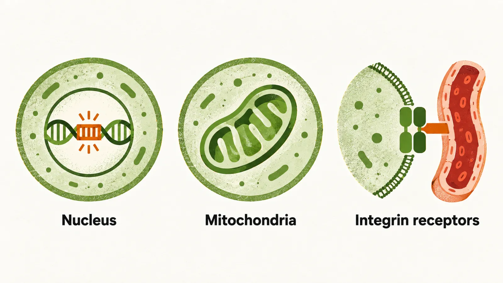
The nuclear and mitochondrial routes use the hormone inside the cell, whereas the integrin route works from the cell membrane. In the nucleus the hormone binds one of two receptors, thyroid receptor α (TRα) and TRβ. TRβ is primarily expressed in brain and liver, and one of its subtypes, TRβ-2, is responsible for the negative feedback on the hypothalamus and the thyrotropic cells.
A genetic condition shows what happens when this receptor fails: in resistance to thyroid hormone (RTH), an autosomal dominant mutation reduces the activity of the TRβ receptor. In these patients there is a high level of unbound thyroid hormone without any increase in the TSH level — a state called euthyroid hyperthyroxinemia. What the hormone then does in the tissues, in development and in adult life, follows from these actions.