
Neoplastic epidural spinal cord compression (ESCC) means a tumor compressing the spinal cord from the epidural space, the compartment that lies between the cord and the bone of the vertebral column. It is an oncological emergency, because cord function that has been lost is often not recovered; the aim is to recognize the compression before the neurological deficit becomes fixed. Why a mass in that space is so dangerous becomes clear from the anatomy.
The cord, its coverings and the epidural space
The spinal cord lies inside the vertebral foramen, the canal formed by the vertebral column. It extends from the foramen magnum at the base of the skull and ends, in most people, at the level of the first or second lumbar vertebra (L1–L2).
Within the canal the cord lies posterior to the vertebral bodies. Between the cord and the bone are three layers of meninges:
- the dura mater, the outermost and toughest layer;
- the arachnoid mater, the middle layer;
- the pia mater, which is applied to the surface of the cord.
Between and around these layers lie the meningeal spaces:
- the epidural space, outside the dura, which contains fat tissue;
- the subdural space, a potential space;
- the subarachnoid space, which contains the cerebrospinal fluid.
The epidural space is the one that matters in this emergency. Because it is bounded on its outer side by bone, a tumor growing into it has nowhere to expand except inward, against the dura and the cord. The mass also obstructs the veins that drain the cord, so edema and reduced blood flow are added to the direct mechanical pressure. That combination is why a relatively small mass can produce symptoms out of proportion to its size, and why reducing the edema is part of treatment.
Causes and mechanisms
ESCC is a final common pathway of several tumors rather than a single disease, and the term covers both solid tumors and hematological malignancies. Apart from the blood cancers, ESCC arises most often by metastasis from solid tumors such as prostate cancer, breast cancer and lung cancer. Among the hematological malignancies, multiple myeloma is the classic cause, because it produces lytic bone lesions that can extend into the canal; lymphoma can also compress the cord.
A tumor reaches the epidural space by one of three routes:
- metastasis from a solid tumor elsewhere, usually to a vertebral body and from there backward into the canal;
- a primary bone-marrow malignancy arising within the spinal column;
- a para-spinal mass that enters the canal through a neural foramen, following the nerve root.
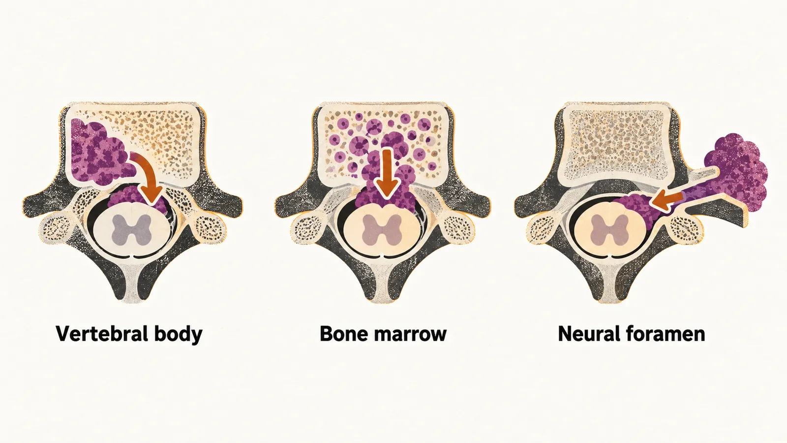
ESCC occurs in about 2.5–5% of patients with a neoplasm, and in about 20% of patients who present with ESCC it is the first sign of an undiagnosed cancer.
Presentation
Pain is the first and most common symptom, present in more than 90% of cases. It begins as an isolated pain without any neurological involvement; on average, neurological involvement appears about 7 weeks later. Because the pain comes first, that interval is the window in which the diagnosis can still be made before cord function is lost.
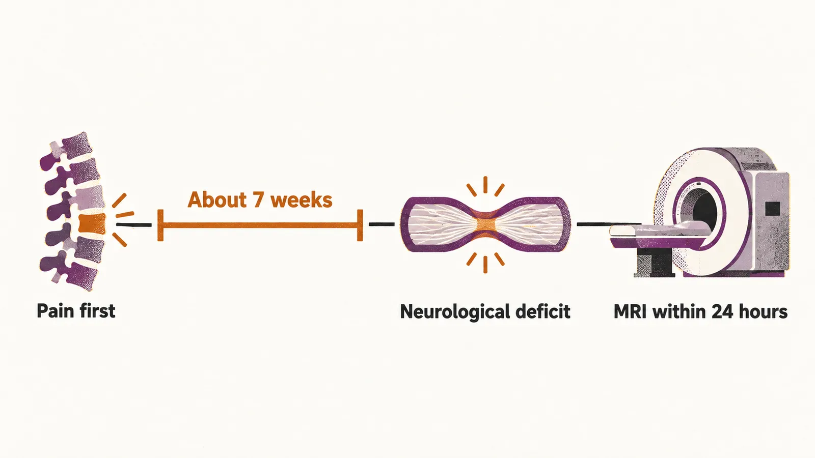
Neurological deficits appear below the level of the compression, because the tumor presses on the cord from outside:
- motor involvement, seen in about 70% of cases, follows the pattern of the dermatomes;
- sensory involvement is not as common as motor involvement and can take the form of ascending paralysis and numbness;
- bladder and bowel dysfunction is seen in about 50% of cases;
- gait ataxia is also a manifestation.
Diagnosis
MRI is the preferred modality, because it shows the cord, the epidural mass and the surrounding soft tissue, and it should be done as soon as possible and always within 24 hours when ESCC is suspected. CT is not as sensitive as MRI. A plain X-ray is often the first radiological investigation, but a positive predictive value of about 80% means that both false positives and false negatives are common, so a normal radiograph does not exclude compression.
Differential diagnosis
The differential matters because the same symptoms are produced by far more common conditions, and a patient with known cancer is easily treated for a musculoskeletal complaint while the cord is being compressed. The conditions to separate from ESCC are musculoskeletal disorders such as muscle spasm, disc herniation and spinal stenosis; a spinal epidural abscess or osteomyelitis (infection of the bone); and metastatic disease of the vertebral column without epidural involvement, which causes pain but no neurological symptoms.
Treatment
When neoplastic ESCC is suspected, high-dose dexamethasone is used to diminish the edema at the site of the lesion. Lowering the edema reduces the external pressure on the spinal cord and the spinal roots, which relieves the neurological symptoms and the pain. The regimen is 16 mg of oral dexamethasone, or an equivalent parenteral dose, given as soon as possible, continued daily while the patient waits for definitive treatment, and then reduced gradually after surgery or at the start of radiotherapy.
Steroids buy time; they do not remove the tumor. Metastatic spinal cord compression is an oncological emergency, and any person with current or previous cancer and symptoms or signs of cord compression needs same-day emergency assessment. Definitive treatment is decompression, radiotherapy or both: surgery is offered when the patient’s performance status allows, usually followed by radiotherapy, while radiosensitive tumors can be treated with radiotherapy alone.
One exception is worth remembering in hematological disease. When lymphoma or myeloma is suspected radiologically but there are no neurological signs or symptoms, corticosteroids are not started before specialist hematological advice, because they can obscure the histological diagnosis of lymphoma.
Compression by a mass, and the steroid caution that goes with lymphoma, come from what a tumor does to its surroundings. Other emergencies of blood cancer arise instead from the biology of the leukemic cell itself, as in acute promyelocytic leukemia, where the danger is bleeding.