
Lymphadenopathy is the enlargement of one or more lymph nodes. Because a lymph node is a filter placed on the course of a lymphatic vessel, how an enlarged node is assessed follows from how the node is built and from the region whose lymph it receives.
The structure of a lymph node
A lymph node is a small, bean-shaped organ that sits on a lymphatic vessel and is organized around the one-way flow of lymph. Lymph — the fluid that drains from the tissues — arrives through several afferent lymphatic vessels that pierce the convex capsule around the surface of the node. It filters through the node and leaves through a single efferent lymphatic vessel at the hilum, the indentation on one side. The same hilum carries the artery and vein that supply the node. Because flow runs in one direction, from capsule to hilum, a microbe or a cell carried in the lymph from a region passes through the node before it can go further.
Inside, the node is divided into a cortex and a medulla. The cortex lies just beneath the capsule and is the B-cell area, containing rounded aggregates called follicles. A primary follicle is a ball of resting B cells that have not yet met their antigen; when a B cell in a follicle is activated, the follicle acquires a pale germinal center and becomes a secondary follicle, the site of the proliferation and terminal differentiation that produce antibody-secreting plasma cells and memory B cells. Deep to the follicles lies the paracortex, the T-cell area, where T cells wait and meet the antigen-presenting cells arriving with the lymph. The medulla is the inner region, carrying the sinuses and cords through which lymph and antibody leave toward the hilum.
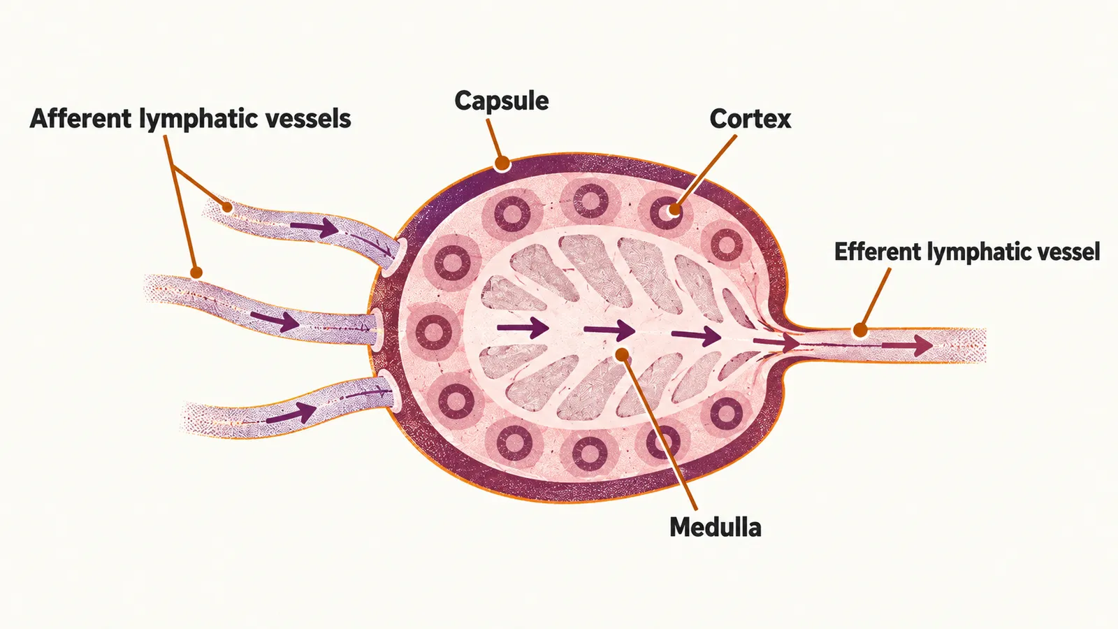
This arrangement explains why the same node enlarges in different ways. When an infection drains to it, the paracortex and follicles fill with activated lymphocytes, so the node becomes soft, tender and mobile. When a malignant clone settles in one of the compartments, the node grows regardless of infection and becomes hard, non-tender and fixed to the tissue around it.
Localized and generalized lymphadenopathy
When the history and the examination do not reveal the cause, the first step is to classify the lymphadenopathy as localized (confined to one region) or generalized (two or more regions involved, which usually points to a systemic disease). Each group has infectious and non-infectious causes, and localized disease is worked up by following the drainage region of the affected nodes.
Malignancy is the exception rather than the rule. Among patients seen in primary care with unexplained lymphadenopathy, only about 1.1% are eventually found to have cancer, and the proportion rises with age — about 4% in patients older than 40 years against 0.4% in those younger than 40.
History and physical examination
The history looks for the exposures and symptoms that point to particular causes. Age and how long the nodes have been enlarged set the background. Contact with animals or eating raw meat, travel and high-risk behavior are the exposures to ask about, and fever, weight loss, night sweats and pruritus are the symptoms. The past medical history and the current drugs complete the picture, since drugs can themselves cause lymphadenopathy.
On examination the first question is again localized or generalized. For the nodes themselves, the features that matter are size, consistency and whether they are mobile or fixed. Associated findings matter as much: a source of infection in the drainage area, or systemic signs such as hepatosplenomegaly or arthralgia.
Supraclavicular lymphadenopathy
Supraclavicular nodes deserve separate mention because they carry the highest risk of malignancy of any peripheral site — biopsy series find cancer in 54% to 85% of them, and the risk is about 90% above the age of 40 years against about 25% below it. What they drain explains why. The right supraclavicular nodes receive lymph from the mediastinum, lungs and esophagus, while the left nodes, known as Virchow’s node, lie at the end of the thoracic duct, which carries lymph from the abdomen. An enlarged left supraclavicular node can therefore be the first sign of a cancer of the stomach, pancreas or another abdominal organ, and supraclavicular nodes are investigated even in a child.
Laboratory testing and imaging
Blood tests come first, because they assess both the marrow and the systemic response. A complete blood count that shows anemia and thrombocytopenia points toward a bone-marrow disorder, while neutrophilia or lymphocytosis points toward an infection, and inflammatory markers such as the erythrocyte sedimentation rate and liver biochemistry add supporting information. A peripheral blood smear is examined for abnormal cells. When malignancy is suspected, the decisive test is biopsy of the node, with immunohistochemistry or flow cytometry used to identify the cell population.
Imaging serves to find nodes that cannot be felt and to look for a primary tumor. Ultrasonography is the initial imaging study for a neck mass in children up to 14 years, and computed tomography for older children and adults. When malignancy is suspected, however, imaging should not delay the biopsy that will make the diagnosis.
When lymphadenopathy needs urgent assessment
Most lymphadenopathy is not an emergency and resolves with the infection that caused it. The features that make malignancy a concern and call for prompt assessment are old age; nodes that are non-tender, non-mobile and firm; and unexplained weight loss.
Management
Management follows the level of concern. When no feature of serious disease is present, the reasonable step is to observe the patient for 3 to 4 weeks: a reactive node usually regresses, and one that does not is then biopsied. When features of malignancy or serious illness are present, biopsy is the next step rather than a period of observation, and the underlying disease is then treated.
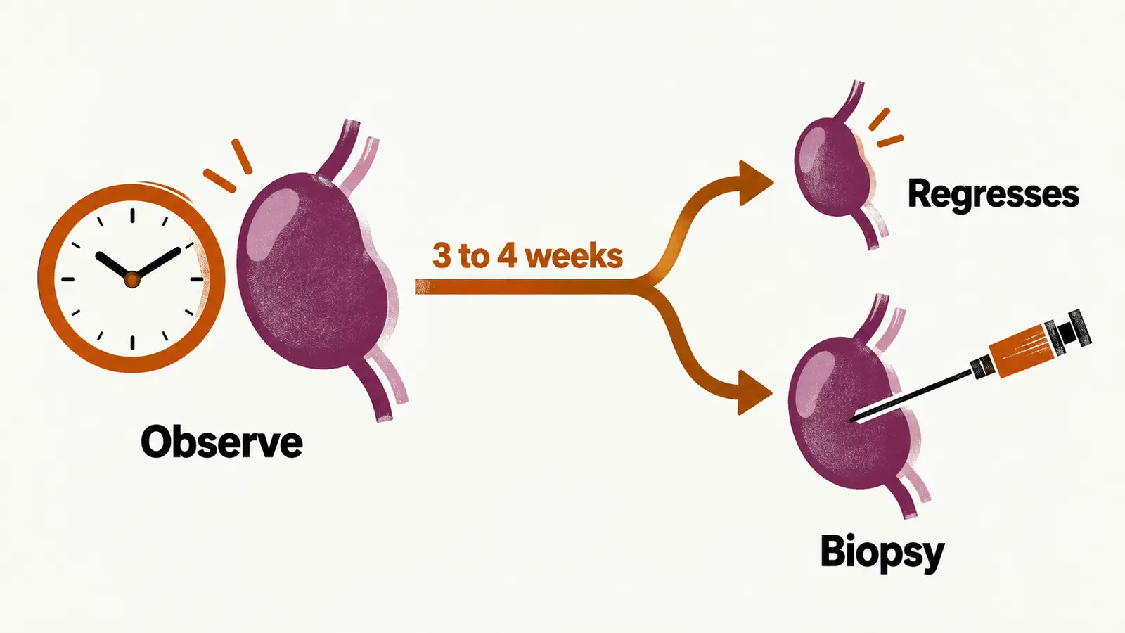
Two cautions apply. Corticosteroids should not be given before a diagnosis is made, because they can shrink a lymphoma and obscure the histology that the biopsy is meant to show. And in acute unilateral cervical lymphadenitis in children with systemic symptoms, empirical antibiotics covering Staphylococcus aureus and group A streptococci may be given.
When the biopsy shows a lymphoid malignancy, the cell population identified is best understood through the stage of B-cell maturation at which it arose.