
The spleen is the largest organ of the lymphatic system and, like the lymph node, a secondary lymphoid tissue. It filters blood rather than lymph, and what it does sets the background for the many conditions that enlarge it.
What the spleen does
The spleen contains two tissues with different jobs. The white pulp is lymphoid tissue arranged around the small arteries; it is a site of innate and adaptive immune responses, of antigen presentation, of terminal B-cell differentiation and of antibody production — the same immune work the lymph node does, but directed at antigens carried in the blood. The red pulp filters the blood: it removes aging or abnormal red cells and destroys cells that antibody has coated, the opsonized cells. The spleen also stores platelets and red cells.
Under abnormal conditions the spleen takes on a third job: when the bone marrow cannot keep up, it becomes a site of extramedullary hematopoiesis, producing blood cells outside the marrow. Because it sequesters platelets, an enlarged spleen can hold a large share of the platelet mass and lower the circulating platelet count, which is why the spleen has to be considered in some thrombotic disorders.
How the spleen enlarges
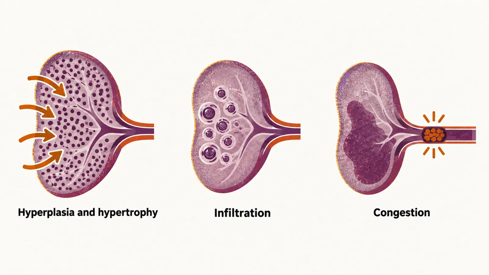
Three mechanisms enlarge the spleen, and they organize its long list of causes. Hyperplasia and hypertrophy are increased function: an infection or a hemolytic anemia makes the spleen work harder, and it grows. Infiltration is the accumulation of abnormal cells — malignant, benign, or the stored material of a storage disorder. Congestion is the pooling of blood when venous outflow is blocked. Some of these are transient: with blood loss, infection or pregnancy the spleen enlarges and then returns to normal when the condition resolves.
Physical examination of the spleen
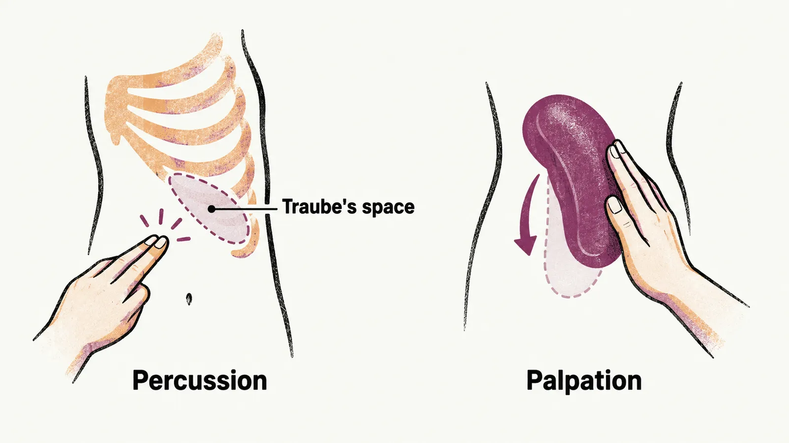
The normal spleen lies almost entirely within the rib cage, and it cannot be palpated unless it has enlarged by at least 40%. Both bedside techniques depend on this, so neither can detect a normal spleen. Percussion of Traube’s space — the area over the spleen bounded above by the sixth rib, laterally by the midaxillary line, and below by the left costal margin — and palpation of the descending spleen on inspiration are the bedside tests. When either is positive, the spleen is enlarged, but the examination is more specific than sensitive, so a negative examination does not exclude splenomegaly and imaging is needed when suspicion remains. Up to 16% of palpable spleens turn out to be normal in size on imaging, so ultrasonography is used to confirm the finding.
Common causes of splenomegaly
In the United States the most common causes of splenomegaly are chronic liver disease, malignancy and infection. Four groups of causes cover these and the others, and each follows from one of the mechanisms above.
Congestive causes raise the pressure in the splenic vein so that blood pools in the spleen; the examples are cirrhosis with portal hypertension, and heart failure.
Neoplastic causes are leukemia and lymphoma infiltrating the spleen; among them, chronic myeloid leukemia is the classic cause of massive splenomegaly.
Infectious causes act through reactive hyperplasia. They include viral infection with EBV (Epstein-Barr virus) or CMV (cytomegalovirus), as in infectious mononucleosis, and bacterial infection with urease-positive organisms such as Brucella; in tropical regions, malaria and schistosomiasis are common causes.
Hematologic causes are the hemolytic anemias, particularly with extravascular hemolysis as in AIHA (autoimmune hemolytic anemia), which make the spleen work harder.
Diagnosis
Splenomegaly is confirmed with abdominal ultrasonography, the first-line imaging study. A splenic diameter larger than 10 cm is generally considered enlarged, and the enlargement is called massive when the lower pole reaches the left lower quadrant or crosses to the right side of the abdomen. Blood tests — a complete blood count, a complete metabolic panel and, when mononucleosis is suspected, a monospot test — screen the hematologic and hepatic causes, and a peripheral blood smear is examined when a hematologic abnormality is found. Further testing is guided by the suspected cause.
Management
Management treats the underlying disease. Spleen-reduction therapy and splenectomy are used for painful or massive splenomegaly, in malignancy, and to control the cytopenias of hypersplenism. Any patient whose splenic function is reduced needs vaccination against encapsulated organisms — pneumococcus, meningococcus and Haemophilus influenzae type b — and against influenza, together with antibiotic prophylaxis; a fever in an asplenic patient is treated as an emergency, because sepsis can progress faster than in a person with a working spleen.
Complications
The most common complications are infection, anemia and splenic rupture. Any trauma to an enlarged spleen can rupture it, and up to 1 in 200 cases of infectious mononucleosis is complicated by rupture, so contact sports are avoided for at least 21 days after the onset of symptoms. Splenic rupture also occurs without trauma in patients with underlying splenic disease, most often malignancy or infection.