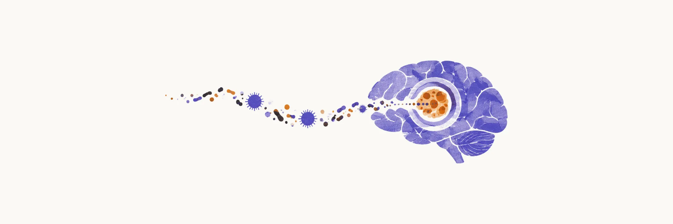
A brain abscess is a localised collection of pus within the parenchyma, the brain tissue itself, walled off by granulation tissue and gliosis (the reactive tissue that forms a capsule around it). It behaves as a space-occupying mass rather than a diffuse inflammation. The clinical signature is the mirror image of meningitis: focal deficits such as hemiparesis or aphasia, with headache and fever, but no meningeal signs. Fever with a focal deficit and no stiff neck should raise abscess before meningitis.
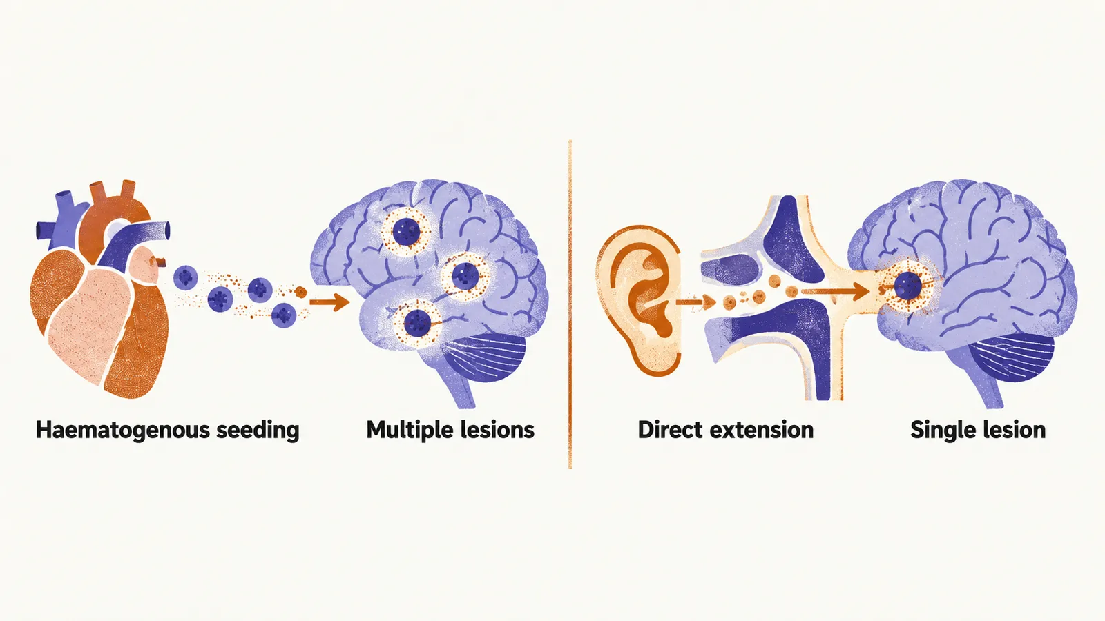
The route by which bacteria arrive shapes where lesions appear. Haematogenous seeding (spread through the blood) from endocarditis, dental, skin, or pulmonary sources lodges organisms in small vessels and favours multiple lesions. Direct extension from otitis media, sinusitis, neurosurgery, or trauma favours a single lesion near the source. Temporal lobe lesions point toward the ear. Frontal lesions point toward the sinuses.
Imaging and the ring-enhancing differential
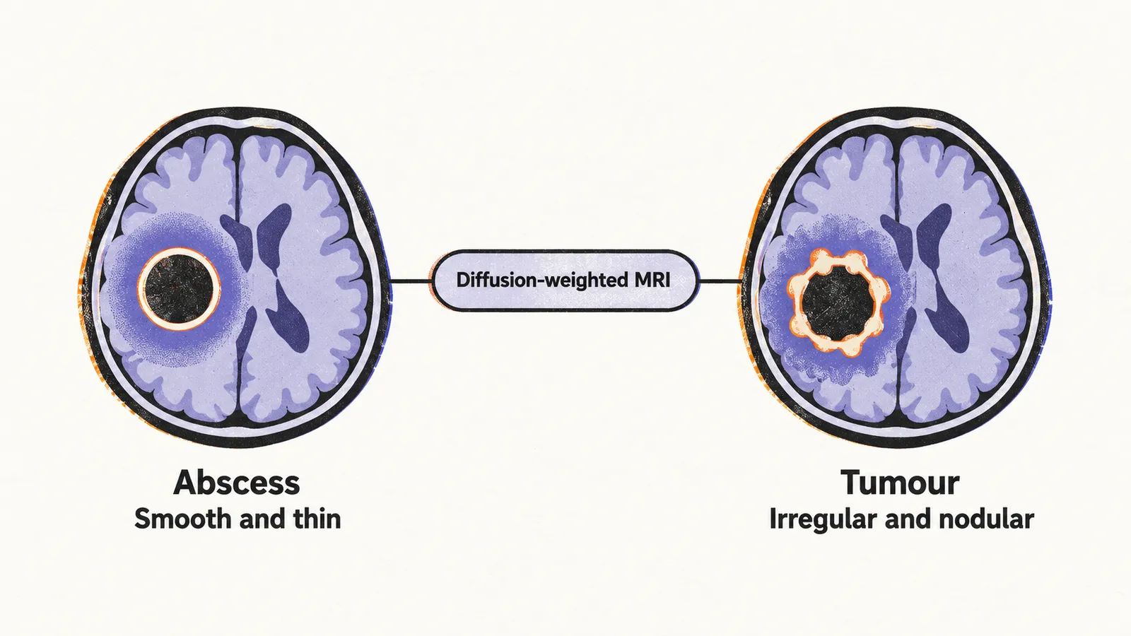
Contrast CT shows the characteristic ring-enhancing lesion: a low-density necrotic centre rimmed by an enhancing inflammatory capsule with surrounding oedema. The differential for such rings includes metastasis, glioblastoma, and toxoplasma lesions in immunocompromised patients. Abscess rings tend to be smooth and thin. Tumour rings tend to be irregular and nodular. Diffusion-weighted MRI is the most reliable separator: the pus cavity restricts diffusion and reads hyperintense, while tumour necrosis generally does not.
Drainage plus antibiotics
Antibiotics alone are insufficient because the capsule blocks penetration and the necrotic core shelters organisms. Needle aspiration or surgical drainage is generally recommended, both to decompress the lesion and to recover the causative pathogen. Lesions above about 2 cm are typically considered for aspiration or excision. Drainage is followed by prolonged targeted antibiotics, typically over many weeks. Empiric cover aims at streptococci, anaerobes, and staphylococci per local protocol until cultures return.
Two safety points matter here. Rupture into the ventricles produces sudden catastrophic ventriculitis (infection of the ventricles) and meningitis and is a surgical emergency. And lumbar puncture is rarely required and must be avoided when abscess is suspected: it is performed only after CT or MRI has excluded dangerous mass effect, because the pressure gradient across the mass risks herniation. Routine cerebrospinal fluid (CSF) analysis is nonspecific in any case.