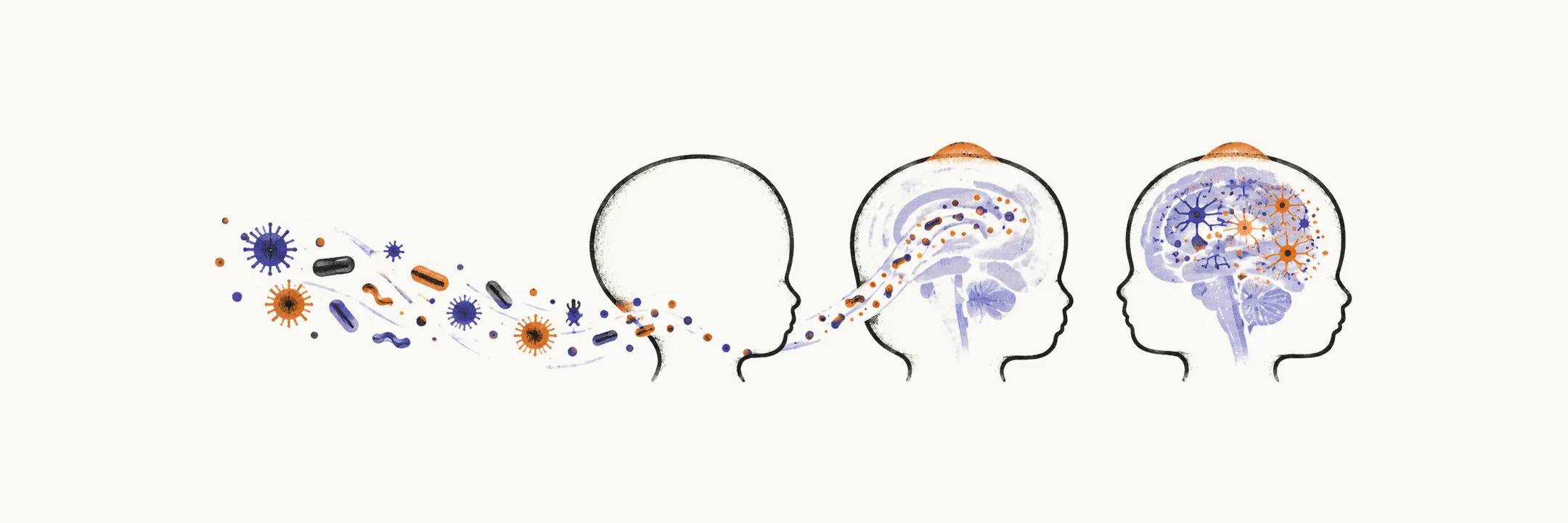
Pediatric CNS infection covers the same anatomy as adult disease: meninges, parenchyma (brain tissue), and focal abscess. Three forces reshape it: an immature immune system, a vaccine history that has rewritten the pathogen landscape, and an examination that grows less specific as the child gets younger. The neonate with meningitis rarely offers neck stiffness. Instead there is poor feeding, lethargy or irritability, apnoea, temperature instability in either direction, and a bulging fontanelle.
The pathogen follows the age
The likely organism is largely predictable from age, and empiric antibiotics (started before the organism is known) follow it. The neonate under 1 month faces group B streptococcus, Escherichia coli, and Listeria from vertical transmission, meaning passage from mother to baby. Group B streptococcus causes over 40% of early-onset neonatal infections, with E. coli next at roughly 30%. Intensive care settings add Klebsiella, staphylococcus, and pseudomonas, with a shift toward resistant and nosocomial (hospital-acquired) organisms in preterm and late-onset disease. From early infancy the world shifts to pneumococcus, meningococcus, and, where unvaccinated, Haemophilus influenzae type b. Past age 5, pneumococcus and meningococcus account for the great majority, and Listeria stops being a routine consideration.
Risk factors modify the map: complement deficiency, crowding, and epidemics for meningococcus; cochlear implants, asplenia, sickle cell disease, HIV, and ear or sinus infection for pneumococcus; immunodeficiency and diabetes for Listeria.
Vaccines changed the epidemiology
Haemophilus type b once caused the great majority of meningitis in unvaccinated under-5s. After conjugate vaccines it has nearly disappeared, with only a small residual from vaccine non-response. Pneumococcal conjugate vaccines cut invasive disease by vaccine serotypes (the strain types a vaccine targets) dramatically. Non-covered serotypes such as 19A then rose to replace them, which is why higher-valency vaccines followed. Meningococcal disease in developed countries is now dominated by group B, which the earliest conjugate vaccines did not cover. The highest incidence sits in infants under 12 months and adolescents around 15 to 19 years. A fully vaccinated schoolchild with bacterial meningitis is therefore more likely to carry a non-vaccine serotype, and the history of doses received changes the differential.
Workup in children
Blood work runs in parallel with the lumbar puncture (the tap, which samples cerebrospinal fluid): cell count with differential, C-reactive protein, procalcitonin, which rises more specifically in bacterial infection, coagulation studies especially when meningococcal disease is possible, simultaneous blood glucose for the fluid ratio, and two blood cultures before antibiotics where feasible without delaying them.
Fluid interpretation mirrors the adult patterns, with one pediatric emphasis: the glucose ratio against simultaneous serum glucose decides, not the absolute number. Viral and bacterial disease cannot be split on clinical grounds, so empiric antimicrobials stand until the cause is confirmed.
Lumbar puncture proceeds without prior CT in most children. Imaging first is reserved for altered consciousness, focal signs, papilledema (swelling of the optic disc), ongoing or recent seizure, evolving purpura (bleeding into the skin), or immunodeficiency. The tap is skipped entirely in shock, respiratory failure, fulminant purpura with instability, intracranial hypertension, profound thrombocytopenia, or local infection. Petechiae (pinpoint skin haemorrhages) with fever and an ill appearance trigger immediate ceftriaxone without waiting for the tap. Polymerase chain reaction (PCR), a test for the organism’s genetic material, rescues the diagnosis when earlier antibiotics have sterilised cultures.
Empiric antibacterial treatment
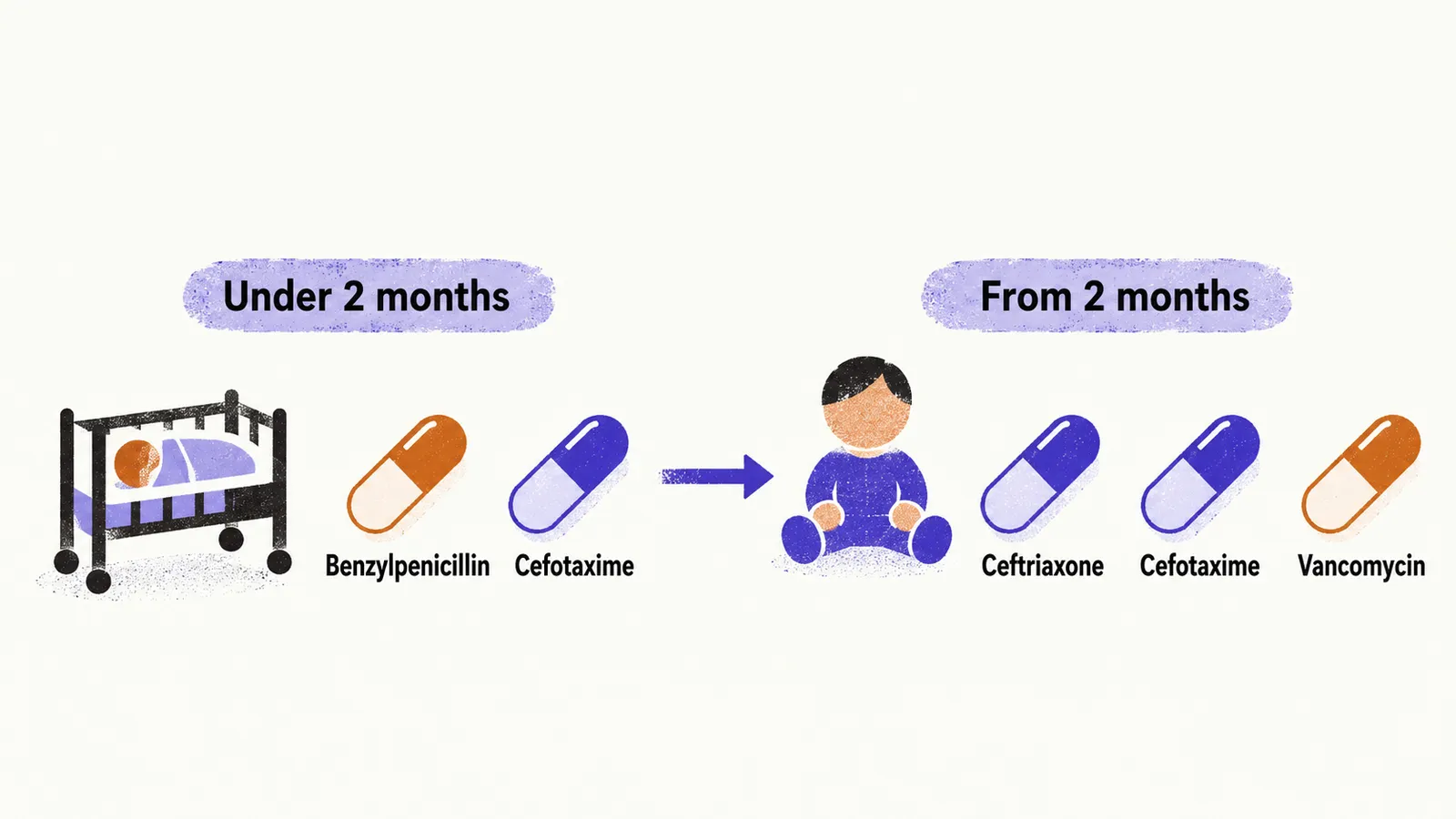
Empiric antibiotics start on suspicion, before results. Suspected early-onset neonatal disease starts ampicillin plus an aminoglycoside or an expanded-spectrum cephalosporin. Ampicillin is non-negotiable because it covers group B streptococcus and Listeria. The age-banded bedside version runs: under 2 months benzylpenicillin plus cefotaxime; from 2 months ceftriaxone or cefotaxime, with vancomycin added when Gram-positive cocci appear. Dexamethasone given with or just before the first antibiotic dose reduces later sequelae over a weight-dosed 4-day course in the older band, notably hearing loss. It is not routine in neonates. Therapy narrows once the organism returns, over roughly 1 to 3 weeks depending on organism and bloodstream involvement.
Herpes encephalitis in children
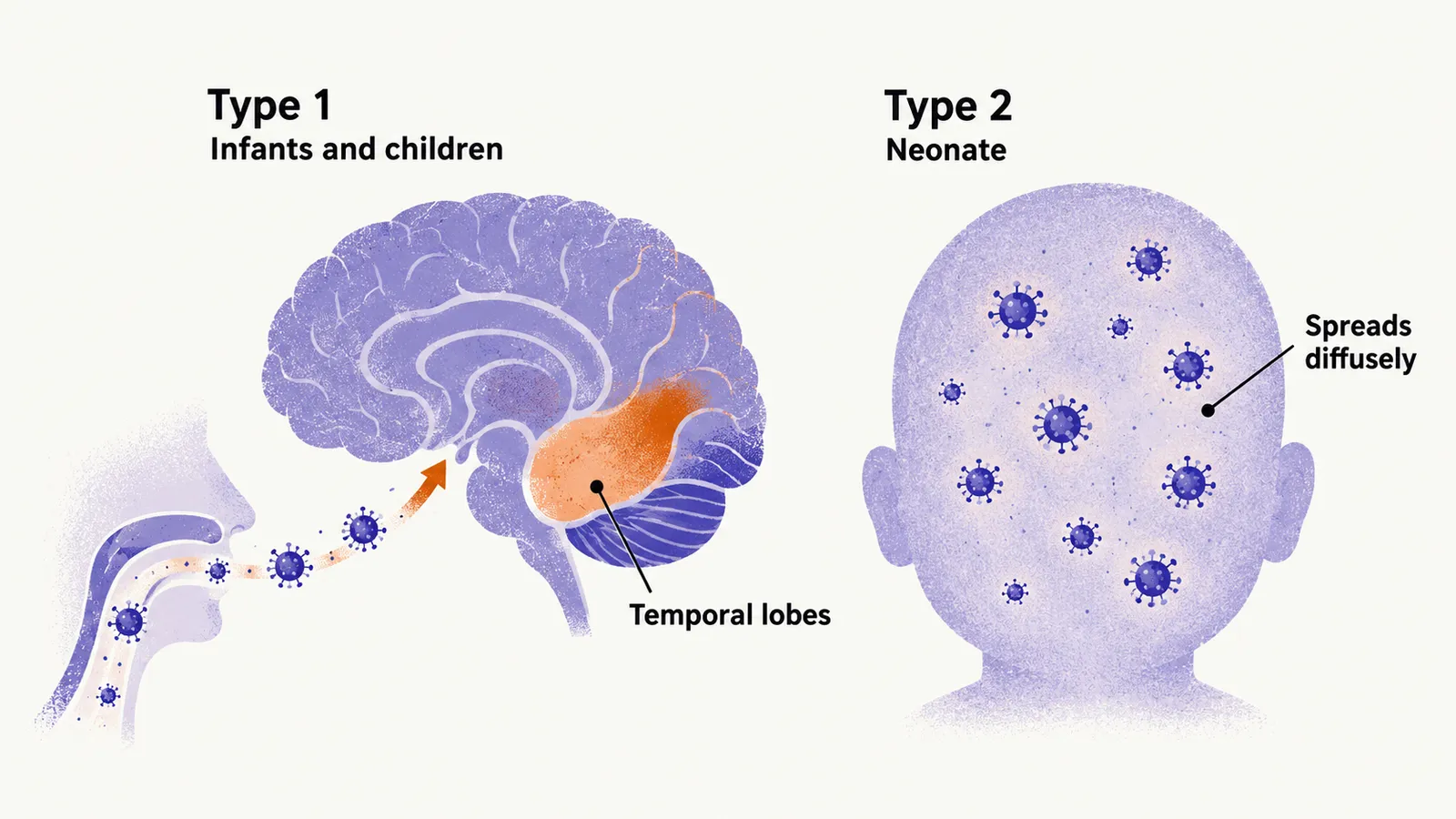
Herpes encephalitis splits by age and route: type 1 reaches infants and children respiratorily and localises to frontal and temporal lobes with fever, headache, psychiatric change, seizures, and focal deficits; type 2 reaches the neonate through the birth canal, spreads diffusely, and declares itself within hours to about 12 days with lethargy, poor feeding, irritability, seizures, and sometimes skin lesions.
Suspected encephalitis gets empiric acyclovir for a full 2 to 3 week course alongside antibacterials until cultures exclude bacterial disease, because missed herpes encephalitis kills and the drug is comparatively safe. Meningitis and encephalitis frequently coexist in children, and meningoencephalitis is often the more honest label.