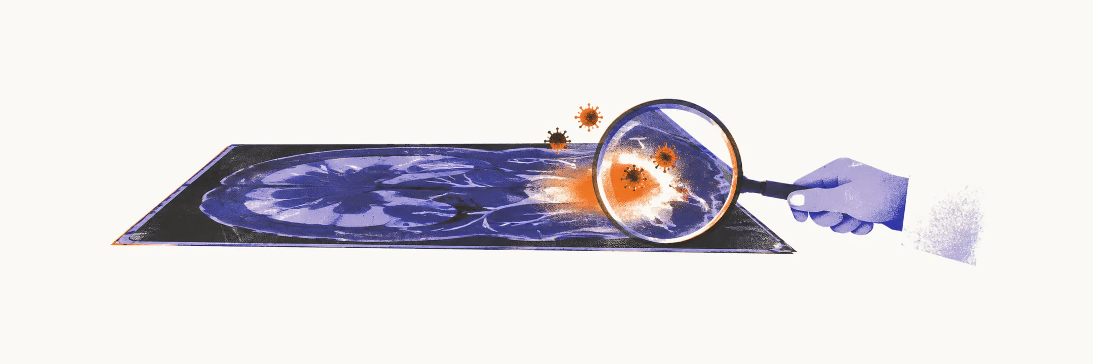
Viral encephalitis is inflammation of the brain parenchyma, the brain tissue itself. Where meningitis gives diffuse meningeal signs, encephalitis gives evidence that brain tissue is malfunctioning: altered mental status, seizures, behavioural change, and focal deficits. Herpes simplex virus type 1 (HSV-1) dominates teaching and practice. It is the most common sporadic cause, it responds to acyclovir, and it is fatal or permanently disabling when missed.
The virus favours the temporal lobes and the adjacent limbic structures such as hippocampus and amygdala. The usual picture is fever with headache progressing over hours to days, then confusion or coma, seizures, personality change, and aphasia or hemiparesis. Because early disease mimics stroke or primary psychiatric illness, the diagnosis is easily delayed.
Virtually all post-neonatal herpes encephalitis is HSV-1. Neonatal disease is more often HSV-2 and spreads more diffusely through the brain.
Diagnosis: MRI, PCR, and the normal-CSF rule
CT is often normal early and never excludes the diagnosis. MRI with T2 and FLAIR sequences is the sensitive study. It shows temporal lobe hyperintensity (bright signal) that is often unilateral or asymmetric. That asymmetry helps against autoimmune limbic encephalitis, in which the immune system attacks the limbic structures and which tends toward bilateral symmetric temporal changes. Imaging alone never settles the cause.
Cerebrospinal fluid (CSF), sampled by lumbar puncture, typically shows lymphocytes, raised protein, and normal glucose, with herpes PCR (polymerase chain reaction, a test for viral genetic material) as the definitive test. PCR for HSV-1 and HSV-2 runs about 96% sensitive and 99% specific. The essential qualifier is that fluid can be entirely normal early on. A single normal lumbar puncture therefore does not overrule a consistent clinical picture. A negative PCR obtained within 3 days of symptom onset in high-probability disease must be repeated, with acyclovir continued meanwhile. The tap and imaging are repeated when suspicion persists.
Acyclovir starts on suspicion
Because early fluid can be entirely normal and an early PCR can be negative, treatment cannot wait for confirmation. Every adult with suspected herpes encephalitis starts weight-based intravenous acyclovir at once, without waiting for PCR. Delay beyond 48 hours tracks with poor outcomes. The course continues for 2 to 3 weeks, with renal monitoring throughout because the drug injures the kidney. The governing rule is to start before confirmation: a few days of unnecessary acyclovir harm little, while a few days of untreated herpes encephalitis destroy brain.
The same drug covers varicella-zoster encephalitis, which is usually less severe. Suspected bacterial meningitis is covered in parallel until cultures return, since the two cannot always be separated at presentation. In children, herpes encephalitis is considered in any child with encephalopathy and in neonates, and empiric antimicrobials (started before the organism is known) are given within 30 minutes of the decision when lumbar puncture must wait.
Other viral causes
Other recognised viral causes include varicella-zoster virus, cytomegalovirus (especially after marrow transplantation), West Nile virus, and enteroviruses. Cytomegalovirus disease is treated with ganciclovir and West Nile disease supportively; exact choices follow local protocols.
Post-infectious encephalitis
Acute disseminated encephalomyelitis is an immune-mediated aftermath of infection or, rarely, vaccination rather than direct viral invasion. It predominantly affects white matter through cross-reactivity with neural antigens. For example, measles causes acute encephalitis in roughly 1 in 1,000 cases, while encephalitis or encephalopathy after measles-containing vaccine is reported in fewer than 1 per million doses.
Because the pathology is immune rather than infectious, steroids rather than antivirals are the treatment. The approach is intravenous steroids followed by an oral taper. IVIG (intravenous immunoglobulin) or plasma exchange is reserved for steroid-refractory cases. There is no standard therapy proven by randomised trials; everything rests on expert opinion and observational series. Most patients recover fully.
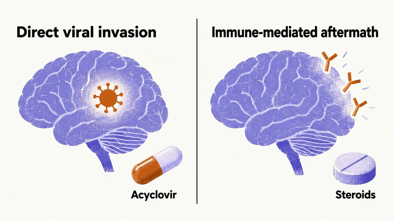
This contrast is the bedside decision the reader must hold: immune aftermath needing steroids versus direct viral invasion needing acyclovir.