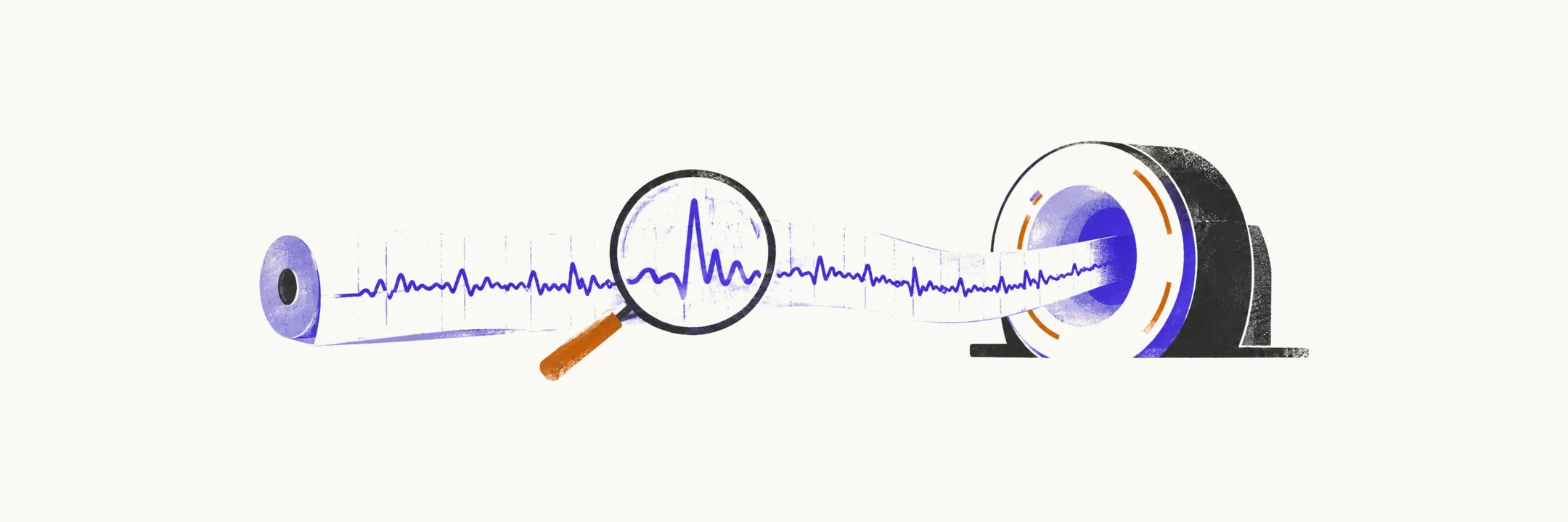
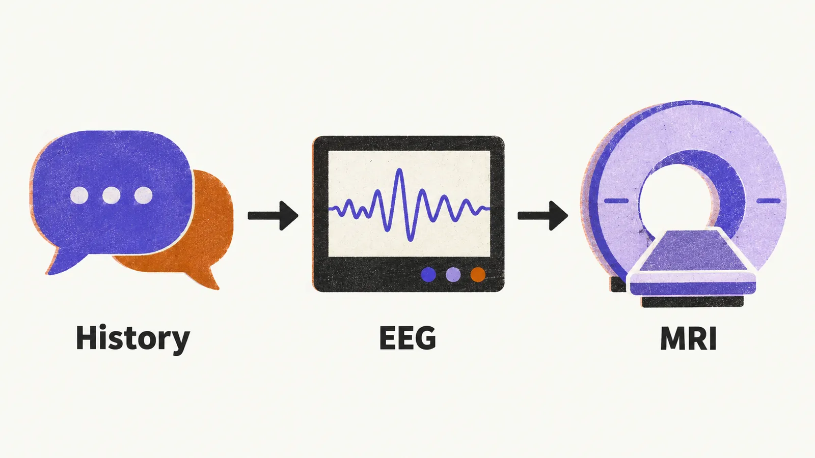
Diagnosing epilepsy is a clinical exercise first and a technical one second. The history decides whether a seizure happened and whether it was epileptic. Investigations then confirm the type and hunt for a cause. Because patients are usually amnestic (unable to remember) for the ictal period, the time of the seizure itself, a witness account is essential. It covers three phases: before the event (triggers, aura, and prodrome, the warning symptoms that precede an attack), during it (motor pattern, awareness, automatisms, cyanosis, incontinence, duration), and after it (the postictal period: confusion, amnesia, sleepiness, tongue biting).
In children this history-taking is the cornerstone, since examination and imaging are normal in the large majority of cases. A normal interictal neurological examination, one done between seizures, is the rule rather than the exception. Abnormalities point toward a structural cause, and a first seizure after middle age should always trigger a search for tumour, metastasis, or stroke.
Electroencephalography
The EEG signal comes from summed postsynaptic potentials of thousands of cortical pyramidal neurons firing together. Only synchronous population activity registers at the scalp. A standard recording lasts about 20 minutes at rest with simple manoeuvres such as eye opening. Two activation procedures, which are provocations meant to bring out discharges, raise the yield. Hyperventilation produces alkalosis and provokes discharges particularly in absence epilepsy. Intermittent photic stimulation uses strobe flashes that provoke photosensitive discharges.
The highest capture probability is within 24 hours of a seizure. A sleep-deprived recording helps when a standard study is negative but suspicion persists. In the emergency department, EEG earns its place in unexplained loss of consciousness and in detecting non-convulsive status epilepticus in comatose patients.
The central finding is the spike: a sharp rapid wave marking synchronous depolarization of a neuronal group. A focal spike points to a localized onset zone. Generalized spikes implicate both hemispheres. The spike-and-wave complex, classically rhythmic at about 3 Hz in absence epilepsy, replaces normal background during the seizure. Isolated spikes and sharp waves, called epileptiform discharges, appear between seizures.
Findings differ by seizure type. Focal seizures show regional spikes and sharp waves over the focus. Generalized seizures show bilateral discharges, often frontal.
Two limits matter. A positive EEG does not always confirm epilepsy, and a negative one never excludes it. About half of children with diagnosed epilepsy have a normal standard recording.
Imaging and other tests
Imaging looks for a structural cause. CT (computed tomography) belongs to the emergency department, where rapid exclusion of haemorrhage or large lesions is the priority. MRI (magnetic resonance imaging) is the outpatient choice because it shows what CT misses: hippocampal sclerosis, cortical malformations, small neoplasms, post-ischaemic and post-traumatic residua, hamartomas, and phakomatoses.
Other tests fill in the picture. Blood tests screen for metabolic and systemic causes. Neuropsychological assessment characterises deficits, especially in temporal lobe epilepsy. PET and SPECT are reserved for the presurgical workup of drug-resistant epilepsy, covered in the nuclear imaging companion hub. Once the event is judged epileptic and its type and cause are clearer, the question becomes whether and how to treat it.