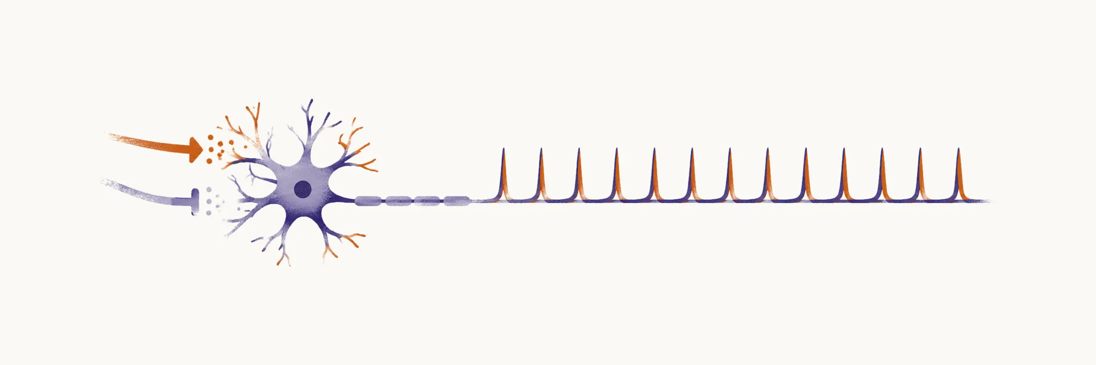
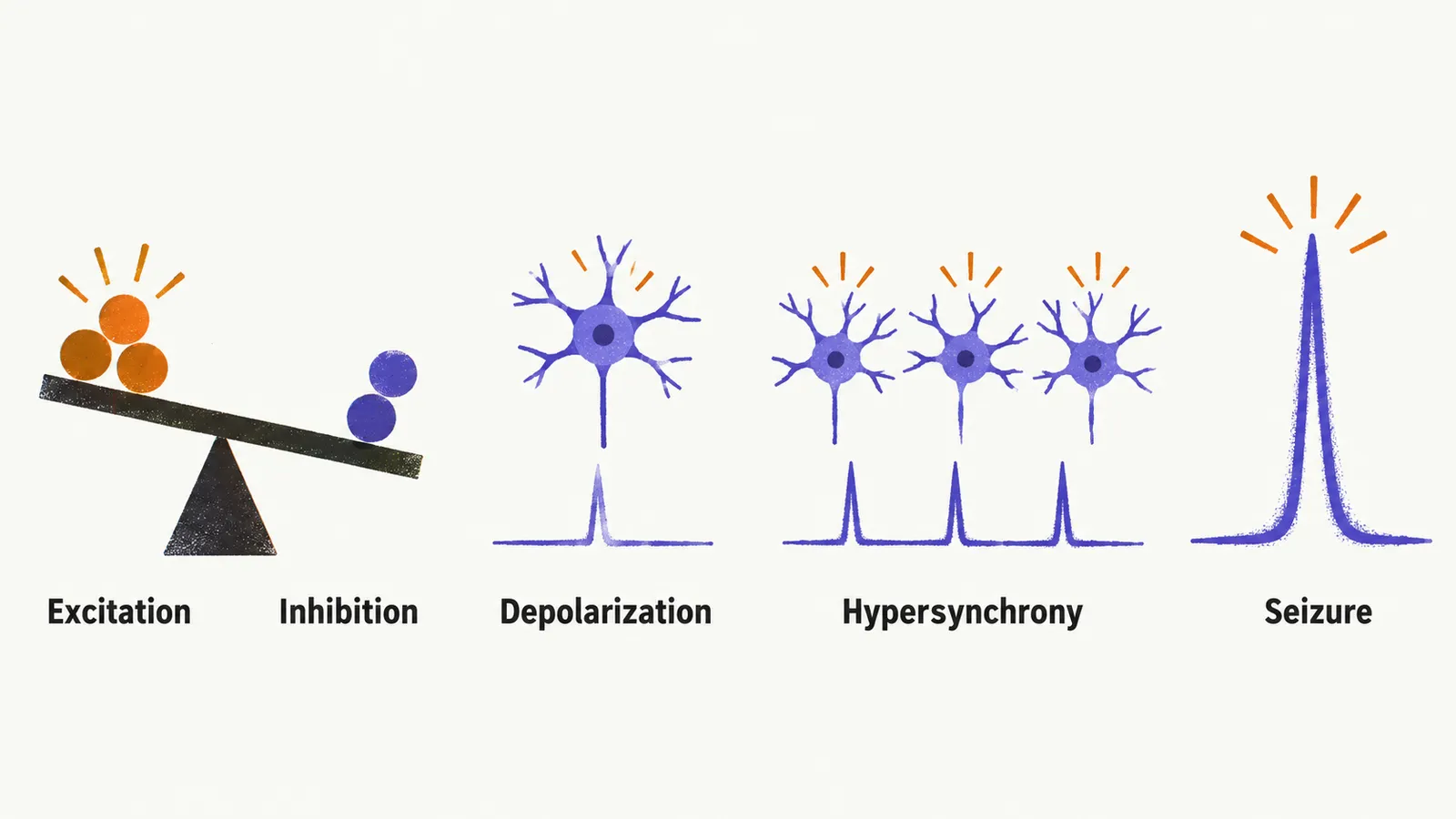
A seizure begins when the balance between excitation and inhibition in cortical networks tips toward runaway excitation. Two pillars carry the process: membrane depolarization and hypersynchrony. The first explains why a neuron fires when it should not. The second explains why that firing becomes a seizure rather than background noise.
Depolarization is a shift in the neuronal membrane potential toward rapid, prolonged, intense firing. The two principal transmitters holding this balance are glutamate, the main excitatory transmitter, and GABA, the main inhibitory one. More glutamatergic drive or less GABAergic restraint makes neurons depolarize more easily. Seizure threshold sits on a continuum. Anyone can seize under extreme provocation such as cocaine intoxication or severe electrolyte disturbance, while people with epilepsy live with an abnormally low threshold because their neurons are hyperexcitable.
Hypersynchrony turns abnormal firing into a seizure. One misfiring neuron produces nothing detectable. A seizure needs thousands of neurons discharging simultaneously enough to summate into a scalp signal. That is what the electroencephalogram (EEG), a scalp recording of cortical electrical activity, registers as spikes and sharp waves. Visible spikes represent only a small fraction of total brain activity, yet their synchronized timing makes them clinically decisive. In generalized seizures the discharge engages both hemispheres rapidly through cortical and subcortical networks. In focal seizures it stays within one hemisphere unless it spreads.
The substrate: channels, pumps, and transmitters
Hyperexcitability has a molecular basis. Voltage-dependent sodium, potassium, and calcium channels can be defective. The membrane ATPase, the enzyme that drives the sodium-potassium pump, can fail. GABA-mediated inhibition can be impaired, and glutamate and aspartate transmission can be heightened. Each shifts the balance toward excitation.
Many genetic epilepsies are channelopathies, disorders of these channels. Sodium channel mutations let neurons fire too easily. Potassium channel mutations prevent clean repolarization, the return of the membrane to its resting potential. Calcium channel mutations drive rhythmic thalamocortical firing in absence seizures.
Structural damage creates the same endpoint by a different road. After a stroke, seizures arise not from dead neurons in the infarct core but from surviving peri-infarct neurons that depolarize more readily, particularly during hyperperfusion, a state of increased blood flow. Roughly 1 in 10 stroke patients develops seizures.
Development adds its own twist. In the immature brain, NMDA-mediated excitation (through the NMDA type of glutamate receptor) is prolonged and GABAergic signalling can be depolarizing rather than hyperpolarizing. This helps explain why seizures are common in children and why most remit as the brain matures. Whatever the route, the end result is abnormal electrical activity, and working out whether a patient has it starts with the history and with recording that activity.