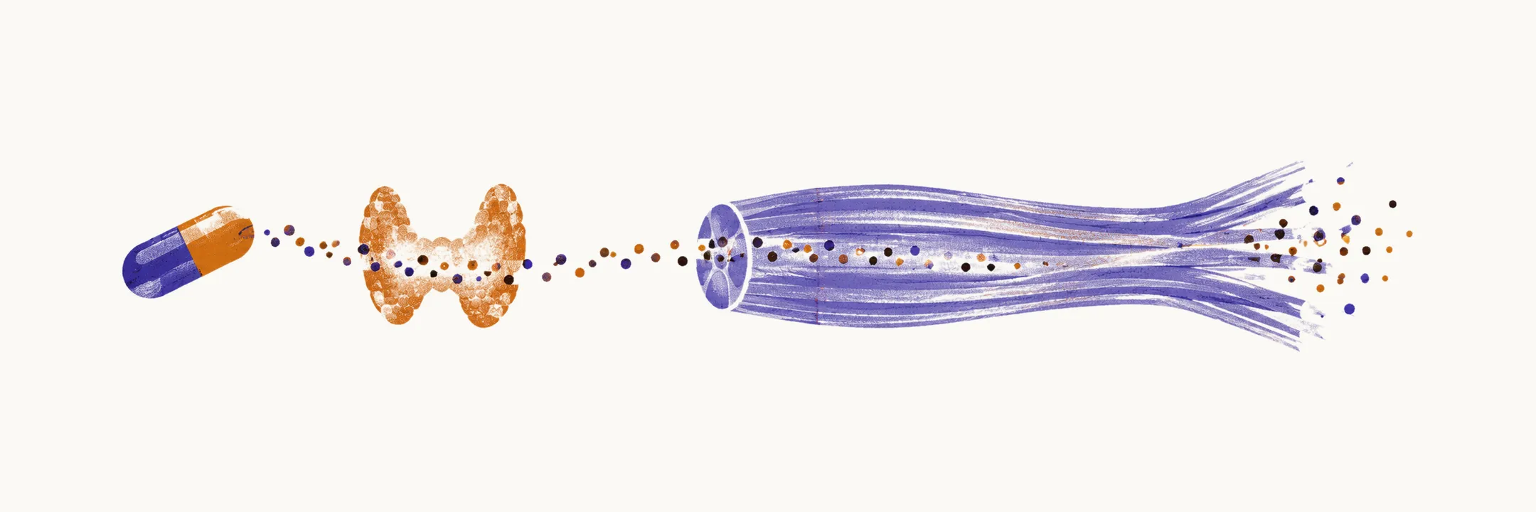
A toxic myopathy is muscle injury caused by a drug or a toxin, and an endocrine myopathy is weakness caused by a hormone disturbance. In both, the muscle is being injured from outside rather than by a defect of its own, which is why the weakness usually reverses once the exposure ends or the hormone balance is corrected. These causes are common enough that they are considered before assuming a primary muscle disease, and a myopathy is occasionally the first sign of the underlying endocrine disease.
Drugs and toxins
Most drug-induced muscle injury is myalgia with a normal or only mildly raised CK, so the diagnosis rests on the temporal relationship with the drug rather than on any laboratory threshold: symptoms typically begin weeks to months after the drug is started and settle within weeks of stopping. A raised CK alone does not establish the diagnosis, and because the differential is broad, endocrine disease, dystrophy, metabolic disease and inflammatory myopathy are excluded alongside it.
Statins are the commonest offender. Muscle symptoms with normal or minimally elevated CK are reported in 7-29% of registry and observational cohorts, whereas statin myopathy with significant CK elevation is rare, at roughly 1 in 1,000 to 1 in 10,000 people on standard doses, and rhabdomyolysis rarer still. Risk rises with interacting drugs such as gemfibrozil, CYP3A4 inhibitors and ciclosporin, and with untreated hypothyroidism. The practical test is the response to withdrawal and to re-challenge with a different statin, and the aim is to keep the patient on the maximum tolerated statin dose with non-statin lipid-lowering agents rather than to abandon lipid control. Weakness with a high CK that does not remit after the statin stops suggests statin-associated immune-mediated necrotising myopathy, which needs immunosuppression rather than further re-challenge.
Glucocorticoids produce a proximal weakness with a normal CK: steroid myopathy is reported in up to half of patients on long-term, high-dose treatment, develops subacutely with atrophy, and shows selective type 2 fibre atrophy on biopsy without inflammation, which separates it from an inflammatory myopathy. The management is to reduce the dose to the lowest effective level, move to alternate-day dosing, or switch to a nonfluorinated steroid, not to add immunosuppression.
Other drugs produce recognisable patterns: colchicine and the antimalarials chloroquine and hydroxychloroquine cause vacuolar or lysosomal change within the fibre, zidovudine injures mitochondria, and alcohol causes both an acute rhabdomyolysis after a heavy binge and a chronic proximal myopathy that improves with abstinence over months to years.
Endocrine causes
Hormone disturbances injure muscle in different ways depending on the gland involved; the thyroid, adrenal and parathyroid glands account for the forms below.
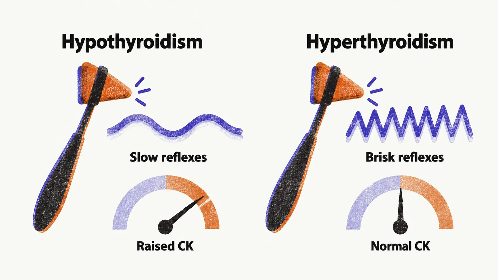
Hypothyroidism is a common cause of a myopathy that can be the presenting problem: neuromuscular symptoms occur in 30-80% of patients, and they can be the first sign of thyroid failure. Proximal weakness, stiffness and cramps are typical, reflexes relax slowly (best tested at the ankle jerk), and percussion can produce myoedema, a localised mounding of the muscle. CK may rise to as much as 10 times normal, and the weakness, the enzyme level and the biopsy changes all improve with levothyroxine replacement.
Hyperthyroidism causes muscle wasting and proximal weakness in about 80% of thyrotoxic patients, with brisk rather than slow reflexes and a CK that is generally normal. Restoring euthyroidism reverses it, and beta-blockade such as propranolol improves muscle function while that is achieved. Thyrotoxicosis can also produce a periodic paralysis, with a low serum potassium during attacks, so thyroid disease belongs in the differential of episodic weakness.
Adrenal causes mirror the steroid effect. Endogenous cortisol excess from Cushing disease or ectopic ACTH (adrenocorticotropic hormone, made outside the pituitary) produces the same proximal weakness and atrophy, with a normal CK and type 2 fibre atrophy on biopsy, and it resolves when the source of excess is removed.
Parathyroid disease changes calcium balance. Hyperparathyroidism causes proximal weakness, wasting, easy fatiguability and hyperreflexia with normal CK and electromyography, and can mimic motor neuron disease when atrophy and spasticity appear; surgery for primary hyperparathyroidism reverses the myopathy. Hypoparathyroidism produces hypocalcaemia, which causes tetany with Chvostek and Trousseau signs rather than fixed weakness.
Removing the cause
These myopathies share one logic: identify the exposure or the hormone disturbance, remove or correct it, and expect improvement over weeks to months. Weakness that continues after the cause has been dealt with is the point at which to revisit the diagnosis.