Diagnosis combines bedside screening, a validated questionnaire, instrumental assessment, and identification of the underlying neurological disease. The goal is to find patients at risk of aspiration — food, liquid or saliva entering the airway — before pneumonia develops. Because patients often do not report symptoms, the sequence starts with simple screens and moves to instrumental tests for those who need them.
Bedside screening
Both bedside screens flag who needs further assessment rather than settling the diagnosis.
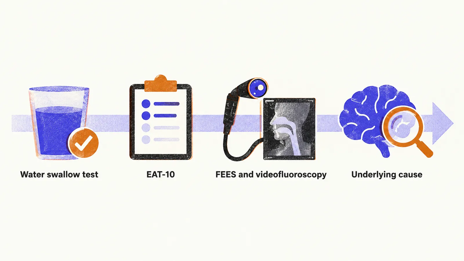
Water swallow test
The patient drinks water — commonly 3 ounces (about 90 ml) without interruption in the Yale protocol — while the observer watches for cough during or after swallowing, a wet voice, throat clearing, or inability to finish the volume. The test is rapid and simple enough for routine use, but it is a screen, not a diagnosis. In validation against endoscopy, the 3-ounce test proved sensitive for aspiration, meaning it is good at picking up patients who aspirate, but with a substantial false-positive rate (specificity around 49%, meaning many patients who do not aspirate still fail). A failed screen therefore needs instrumental confirmation rather than immediate tube feeding.
EAT-10 questionnaire
The Eating Assessment Tool (EAT-10) is a 10-item self-report covering swallowing ability, coughing with meals, and food sticking in the throat, with total scores from 0 to 40. A total score of 3 or higher is considered abnormal and warrants further assessment. The cutoff of 3 is supported by the original validation and by meta-analysis, which found a better balance of sensitivity and specificity there than at lower cutoffs.
Instrumental assessment
Instrumental tests show directly what happens to the bolus during the swallow, which a screen cannot. They are the only way to detect silent aspiration.
FEES (fiberoptic endoscopic evaluation of swallowing)
A flexible endoscope is passed through the nose, and the patient swallows coloured food or liquid while the pharynx is observed. FEES is repeatable at the bedside and shows pharyngeal residue well. Its limits are a brief view loss during the swallow itself (white-out), poor visualisation of the oral phase, and no information about the esophageal phase.
Videofluoroscopy
Real-time X-ray imaging of swallowing with barium-coated foods of different consistencies. It shows oral and pharyngeal dynamics better than endoscopy. The trade-offs are ionising radiation, difficulty for patients with limited mobility, and limited availability.
Pharyngeal manometry and cricopharyngeal EMG
Manometry assesses pharyngeal peristalsis and upper esophageal sphincter function, relevant for example in myotonic dystrophy with incomplete sphincter relaxation. Electromyography of the cricopharyngeal muscle can demonstrate failure of relaxation during swallowing.
Severity scales
Once swallowing has been examined, two scales express how severe the problem is. DOSS grades overall functional severity and what the patient can safely eat, while PAS grades what happens to material in the airway during individual swallows.
DOSS (Dysphagia Outcome and Severity Scale)
DOSS grades functional severity in 7 levels, from 1 (severe: nothing by mouth, alternative feeding required) through intermediate levels needing supervision and texture restriction, to 7 (normal swallowing). In simplified form: levels 6–7 manage a normal diet, levels 3–5 need modified texture and supervision, levels 1–2 need alternative feeding.
PAS (Penetration-Aspiration Scale)
PAS grades each swallow in 8 levels: 1 means nothing enters the airway; 2–5 mean material enters above or at the vocal folds with varying clearance (penetration); 6–8 mean material passes below the folds (aspiration), with level 8 meaning no effort to eject — silent aspiration, the most dangerous grade.
Peak cough flow measurement
The tests above show whether material enters the airway. Whether the patient can then clear it depends on the strength of the cough, which is what peak cough flow measures.
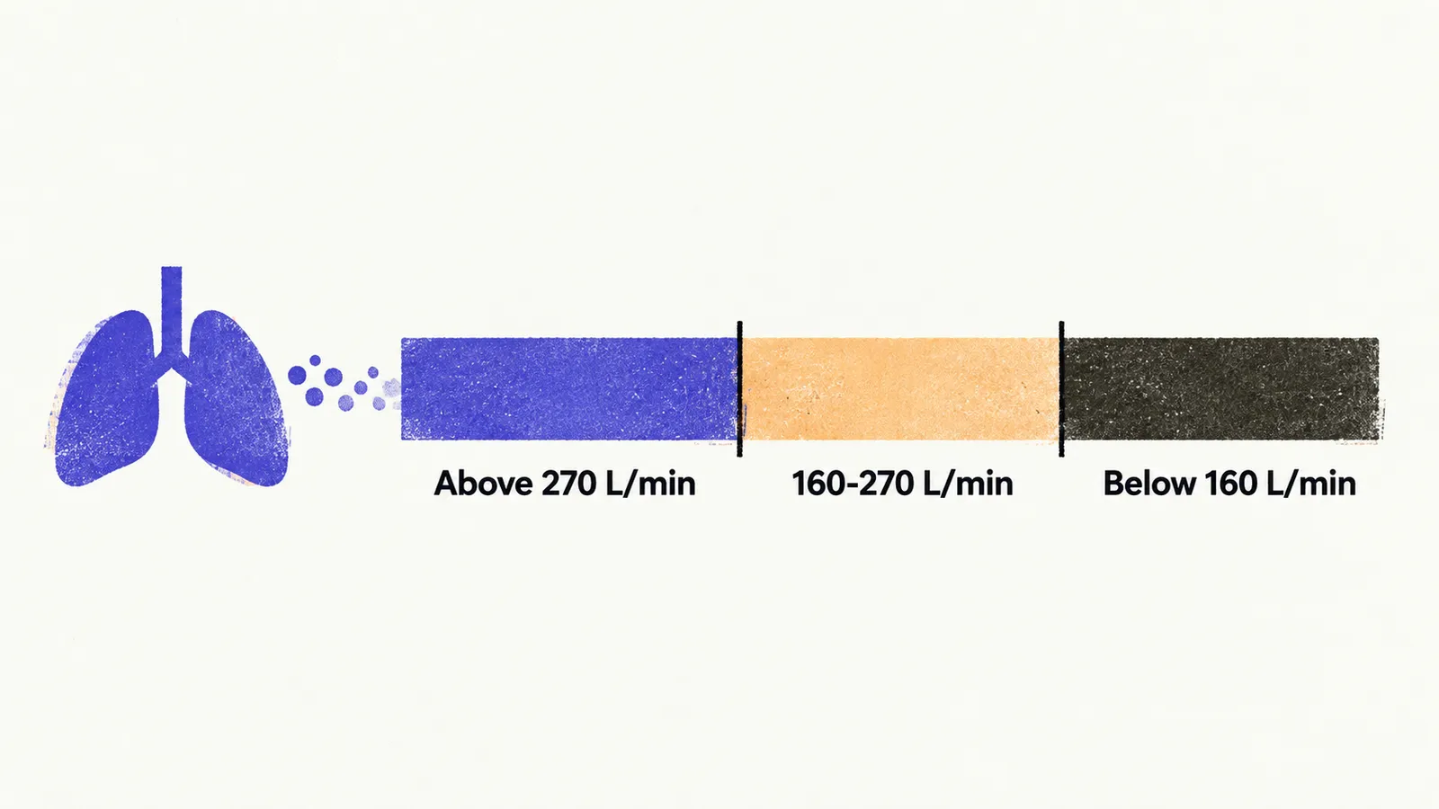
Peak cough flow (PCF), measured in litres per minute with a handheld meter, tests whether the patient can clear what is aspirated:
- Above about 270 L/min: effective cough with adequate reserve.
- Roughly 160–270 L/min: limited reserve, easily exhausted by repeated coughing.
- Below about 160 L/min: ineffective cough that cannot clear secretions, marking the range where assisted-cough techniques are considered.
Exact cutoffs vary somewhat between guidelines; the direction is what matters. In myasthenia gravis, PCF falls with successive efforts — a fatigable pattern that distinguishes it from fixed weakness such as amyotrophic lateral sclerosis (ALS) or stroke, where the low value stays low.
Identifying the underlying neurological cause
Dysphagia is a symptom of many neurological diseases, and the accompanying features point to which one is responsible. The list below gives, for each disease, what characterises it and its link with dysphagia.
- Stroke. Dysphagia affects roughly half of stroke patients. Bilateral opercular infarction (Foix-Chavany-Marie syndrome) abolishes voluntary face, tongue, and pharynx movements while automatic movements survive.
- Lateral medullary (Wallenberg) syndrome. A posterior inferior cerebellar artery infarct producing dysphagia with ipsilateral Horner syndrome, ipsilateral ataxia, contralateral body sensory loss, hoarse voice, and reduced cough reflex.
- Parkinson disease. Dysphagia is reported in a wide range of patients across studies, involving oral, pharyngeal, and esophageal phases, with drooling, delayed gastric emptying, and constipation as associated features.
- ALS. Bulbar symptoms present first in roughly one quarter to one third of patients, and swallowing is affected in nearly all during the disease course, predominantly in the oral phase. Upper and lower motor neuron signs together are the hallmark. Disease from SOD1 mutations is treatable in a small subset with intrathecal antisense therapy.
- Oculopharyngeal muscular dystrophy. Autosomal dominant triplet-repeat disease with bilateral ptosis, dysphagia, and proximal weakness — unexplained dysphagia should prompt a look at the eyes.
- Myotonic dystrophy type 1. The most common adult muscular dystrophy; cohort studies find dysphagia on testing in most patients, largely silent. Incomplete upper sphincter relaxation and esophageal dysmotility are the mechanisms.
- Myasthenia gravis. Dysphagia affects a substantial minority of generalised disease, is especially frequent with anti-MuSK antibodies, and underlies about half of myasthenic crises. Fatigability is the discriminator.
- Multiple sclerosis. Reported in a wide minority of patients at oral, pharyngeal, and esophageal levels; estimates vary with case mix and testing.
- Polymyositis and dermatomyositis. Dysphagia is a negative prognostic indicator, more frequent and severe in dermatomyositis.
Whatever the cause, the dysphagia itself matters through what it leads to: pneumonia, malnutrition, choking, and their effect on outcome.