Nutritional support and respiratory planning must start early in progressive disease — before the patient can no longer tolerate procedures, and before malnutrition and aspiration pneumonia reinforce each other. Delay narrows every option.
Aspiration is dangerous because aspirated material is not always cleared: an ineffective cough, impaired cellular immunity, and reduced mucociliary and lymphatic clearance all allow it to become pneumonia. Nutrition, hydration, oral hygiene, reflux management, and ventilation support act on those same factors, which is why they lower pneumonia risk even when some aspiration continues.
Feeding tubes: plan early
Percutaneous endoscopic gastrostomy (PEG) or jejunostomy (PEJ) placement needs the patient to tolerate lying flat and to have adequate respiratory reserve for the procedure and its sedation. Both decline in progressive neuromuscular disease, so the discussion belongs in advance care planning — framed as possible supplementary feeding if swallowing deteriorates — rather than at the point of crisis.
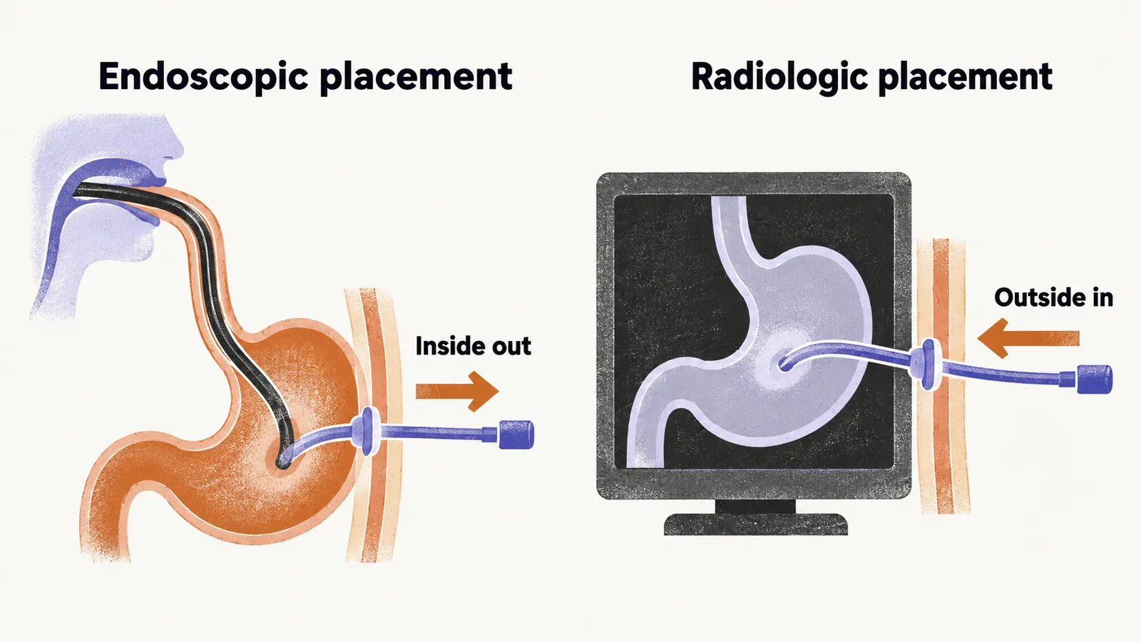
Two placement routes exist. Endoscopic placement passes an endoscope into the stomach and places the tube from inside out; it is the standard approach but needs airway management in an already compromised patient. Radiologic placement inflates the stomach through a nasogastric tube and places the tube from outside in under imaging guidance, which allows ventilation to continue during the procedure — an advantage once the patient needs ventilatory support.
Practical complications include tube dislodgement, for which caregivers must know how to keep the tract open and seek urgent care, since the tract can close quickly; infection; and later exchange for a low-profile button device once the tract matures.
Cough assessment
Cough is the main defence against aspiration, and a cough is a sequence of three steps — a breath in, a build-up of pressure behind a closed glottis, and a forceful expulsion — so it can fail at any one of them in a recognisable way:
| Cough phase | What fails | Example conditions |
|---|---|---|
| Inspiration | Inspiratory muscle weakness limits inhaled volume | Amyotrophic lateral sclerosis (ALS), muscular dystrophy, myasthenia gravis |
| Compression | Glottic closure fails so pressure cannot build | Brainstem stroke, vocal cord paralysis, open tracheostomy |
| Expulsion | Expiratory weakness limits flow | ALS, myasthenia gravis, muscular dystrophy |
How strong the cough is overall can be measured. Peak cough flow, measured in litres per minute, shows whether the patient can clear what is aspirated: above about 270 L/min the cough is effective, roughly 160–270 L/min leaves limited reserve that repeated coughing exhausts, and below about 160 L/min the cough cannot clear secretions.
Do not prescribe mucolytics without assessing cough. Thinning secretions in a patient who cannot cough makes clearance harder, not easier, and can precipitate respiratory failure. The same caution applies after brainstem stroke with impaired cranial nerve function.
Cough-assist machines
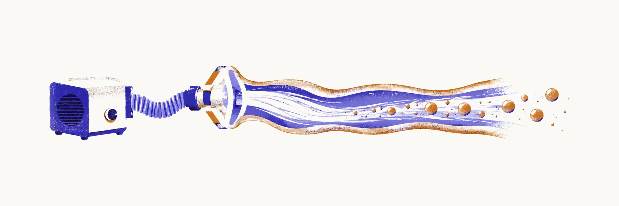
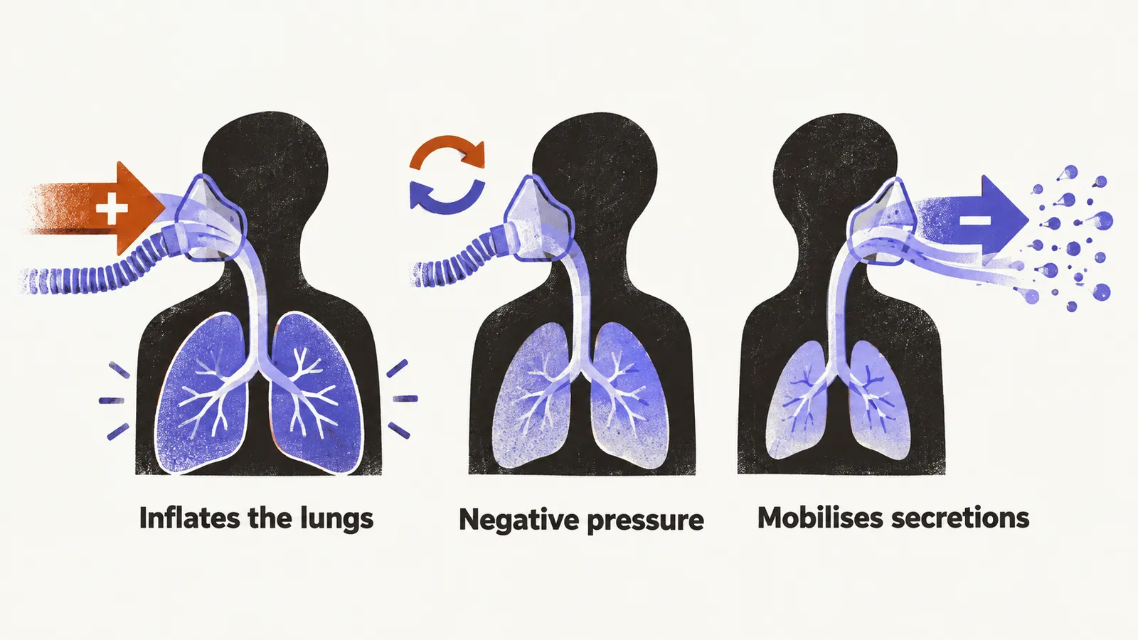
Mechanical insufflation-exsufflation devices mimic the cough cycle: positive pressure inflates the lungs, then rapidly switches to negative pressure to generate a high expiratory flow that mobilises secretions. Full-size devices reach roughly +70 and −70 cmH2O. They are used in neuromuscular weakness with ineffective cough, including ALS, myasthenia gravis, and muscular dystrophy, though some patients cannot tolerate them.
The oxygen hazard in neuromuscular disease
A neuromuscular patient with weak cough accumulates secretions and arrives with low oxygen saturation. The low saturation reflects hypoventilation from muscle weakness, not primary lung disease, and the remaining ventilatory drive may depend substantially on the hypoxic stimulus. Giving supplemental oxygen alone can blunt that drive and precipitate hypercapnic respiratory failure, meaning failure with carbon dioxide retention. The correct response is ventilatory support (such as non-invasive ventilation) combined with secretion clearance, following local protocols — not oxygen alone.
Compensatory postures and strategies
Alongside tube feeding and cough support, several adjustments make the swallow itself safer. They differ in mechanism and in when they apply.
- Chin-tuck: flexing the head forward widens the valleculae, the small pockets between the tongue base and epiglottis, and pushes the tongue base and epiglottis backward, protecting the airway. It suits delayed swallow reflex and spillage before the swallow.
- Positioning: some patients with fixed neck postures swallow more safely in a lateral position.
- Tracheostomy: never eat with the tube open; the cuff should be inflated to limit aspiration past the tube.
- Consistency modification: thicken liquids, avoid mixed textures, and adjust bolus volume and temperature to aid sensory input. Foods that fragment into small sticky pieces — hard bread, crackers, rice, stringy meats and vegetables, whole legumes, stringy cheese — are the usual culprits to avoid.
- Sensory and electrical stimulation of swallowing muscles is described as an adjunct. Reported effects develop over weeks rather than immediately, and detailed protocols and evidence grading are limited, so this remains rehabilitative rather than established therapy.