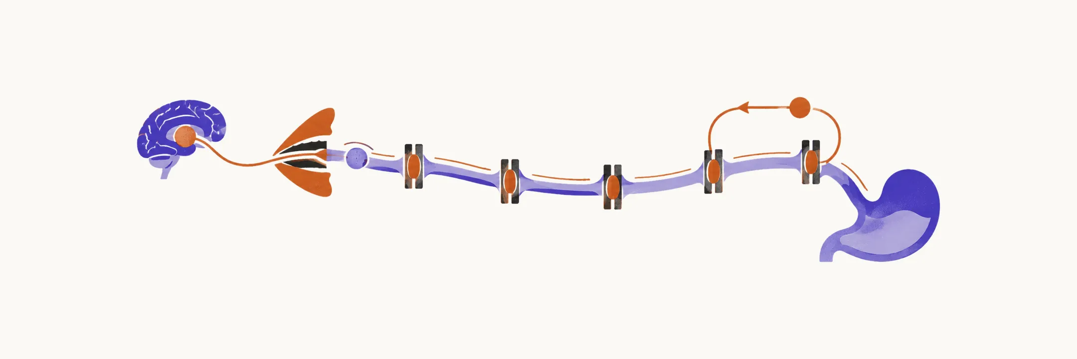
A swallow is not a single reflex. It begins with the decision to eat and ends when peristalsis — the wave of muscle contraction that propels food onward — carries the bolus, the ball of food or liquid being swallowed, from the mouth to the stomach. The nervous system organises that sequence across several levels: cortical regions plan and modulate it, the cerebellum and basal ganglia coordinate it, and brainstem circuits execute the involuntary part. The sequence is partly voluntary and partly automatic, and it is carried out by dozens of muscles supplied by several cranial nerves and cervical roots. A lesion at any level — cortex, brainstem, or peripheral nerve — can therefore produce dysphagia, and the level of the lesion shapes the pattern.
Neural control of swallowing
Cortical regions including the sensorimotor cortex, supplementary motor area, cingulate gyrus, and anterior insula participate in planning and modulation, while the cerebellum and basal ganglia contribute coordination. Brainstem central pattern generators — networks of neurons that produce a stereotyped sequence without needing conscious control — built around the nucleus tractus solitarius and motor cranial nerve nuclei execute the involuntary part of the sequence. Cortical representation is partly asymmetric, which helps explain why some hemispheric strokes impair swallowing more than others.
The muscles themselves are supplied mainly by five cranial nerves, and what each nerve contributes explains what its loss takes away:
| Nerve | Contribution to swallowing | Typical deficit when it fails |
|---|---|---|
| Trigeminal (V) | Sensation of the face and anterior tongue; chewing muscles, mouth floor, soft palate | Poor chewing and mouth-floor support, reduced elevation of the hyoid and larynx |
| Facial (VII) | Lip seal through the orbicularis oris and buccinator; salivary gland innervation | Weak lip closure, anterior spillage, oral residue, dry mouth |
| Glossopharyngeal (IX) | Sensation of the posterior tongue and pharynx; the stylopharyngeus and other muscles that elevate the pharynx and soft palate | Impaired pharyngeal transport and upper sphincter opening, pharyngeal pooling |
| Vagus (X) | Motor supply to the pharynx and larynx; sensory supply of the larynx | Poor closure between the soft palate and pharynx, weak pharyngeal contraction and cough, impaired airway protection |
| Hypoglossal (XII) | All intrinsic and extrinsic tongue muscles except palatoglossus | Weak bolus control and propulsion, early spillage into the pharynx, oral residue |
Cervical roots contribute too: the ansa cervicalis supplies the strap muscles that help elevate the larynx, and losing that supply impairs opening of the upper esophageal sphincter.
The phases of a swallow, and how neurological disease disrupts them
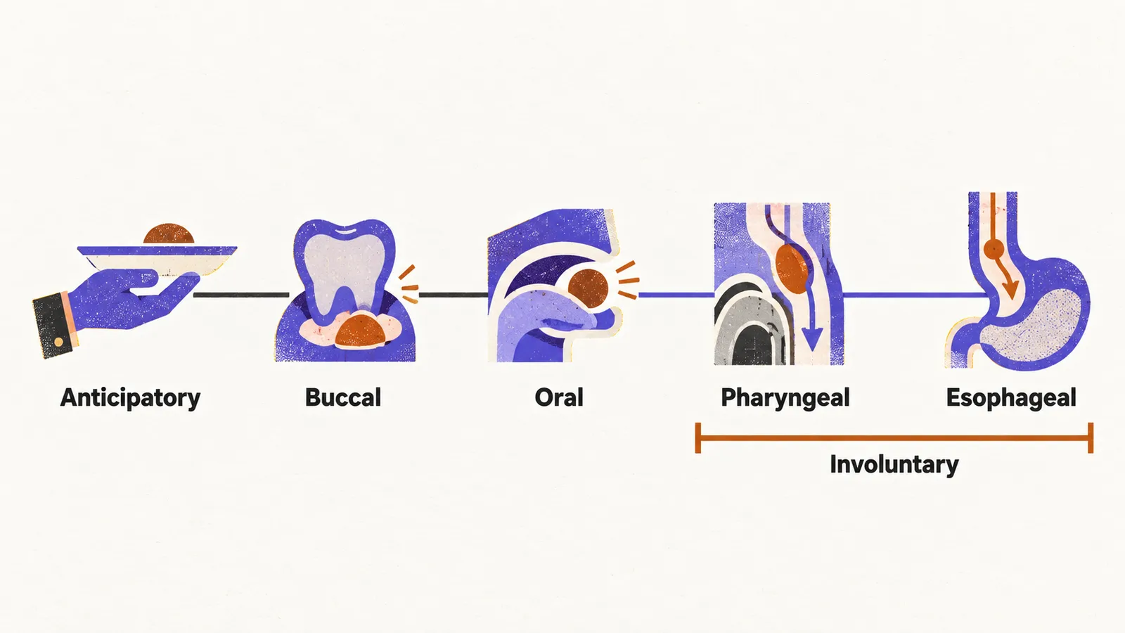
Because the sequence runs from voluntary to involuntary control, the swallow is divided into five phases, each with its own control and its own points of failure. The table sets each phase beside the neurological conditions that typically disrupt it.
| Phase | Control | What happens | What neurological disease disrupts |
|---|---|---|---|
| Anticipatory | Voluntary and cognitive | Bringing food to the mouth, recognising food | Dementia, frontal lesions |
| Buccal (oral preparation) | Voluntary | Chewing, manipulating food, forming the bolus | Facial weakness, bradykinetic (slowed) tongue movement in Parkinson disease |
| Oral | Voluntary (last voluntary phase) | Pushing the bolus past the fauces, the opening from the mouth into the throat | Tongue weakness in amyotrophic lateral sclerosis (ALS) or myasthenia gravis |
| Pharyngeal | Involuntary (first involuntary phase) | The bolus crosses the airway-digestive crossroad while sphincters coordinate | Brainstem stroke, cranial neuropathies |
| Esophageal | Involuntary | Peristalsis carries the bolus to the stomach | Esophageal dysmotility, as in myotonic dystrophy |
Why the level of the lesion shapes the pattern
Cortical lesions tend to interfere most with the parts of swallowing that are under voluntary control: bolus manipulation and chewing in the oral phase, and initiation of the pharyngeal phase. Medullary lesions disturb the patterning of the pharyngeal phase itself, so the pharyngeal component becomes delayed and discoordinated rather than simply weak, and the disturbance can involve both sides. Peripheral lesions, such as a cranial neuropathy or a disorder at the neuromuscular junction, mostly produce weakness of particular muscles, so the deficit follows the muscles involved rather than the level. Cortical asymmetry adds a further variable: the two hemispheres do not contribute equally to swallowing, which helps explain why some hemispheric strokes impair it more than others.
Whatever the level involved, the pattern of failure is what the bedside assessment has to detect, because patients often do not report the problem themselves.