
FDG-PET (positron emission tomography) images brain glucose metabolism with the tracer fluorodeoxyglucose. It does not diagnose epilepsy; electroencephalography (EEG) and history do that. What it solves is the spatial problem EEG cannot: where the seizure focus sits. Between seizures (the interictal state), the cortex surrounding the onset zone shows reduced glucose metabolism, or hypometabolism.
The explanation is inhibitory. The epileptogenic lesion fires continuous pathological activity, and surrounding circuits suppress its spread through inhibition, which consumes less energy, with neuronal loss and reduced synaptic density adding to the deficit. The hypometabolic zone runs wider than the lesion itself but reliably overlaps it, which is why PET lateralizes (says which side) and roughly localizes even when exact margins stay uncertain.
The uptake protocol
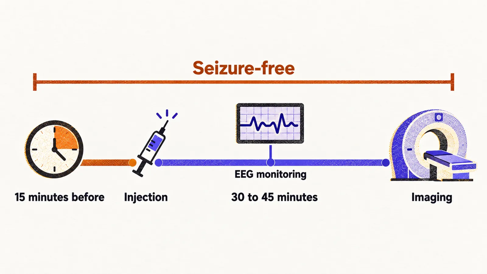
FDG needs 30 to 45 minutes from injection to imaging, and the metabolic state at injection imprints the whole scan. The patient must therefore remain seizure-free throughout uptake, including roughly the 15 minutes before injection, or ictal metabolism (metabolism during a seizure) contaminates the image into an uninterpretable blend. EEG monitoring during uptake is mandatory to certify the interictal state.
Temporal lobe epilepsy
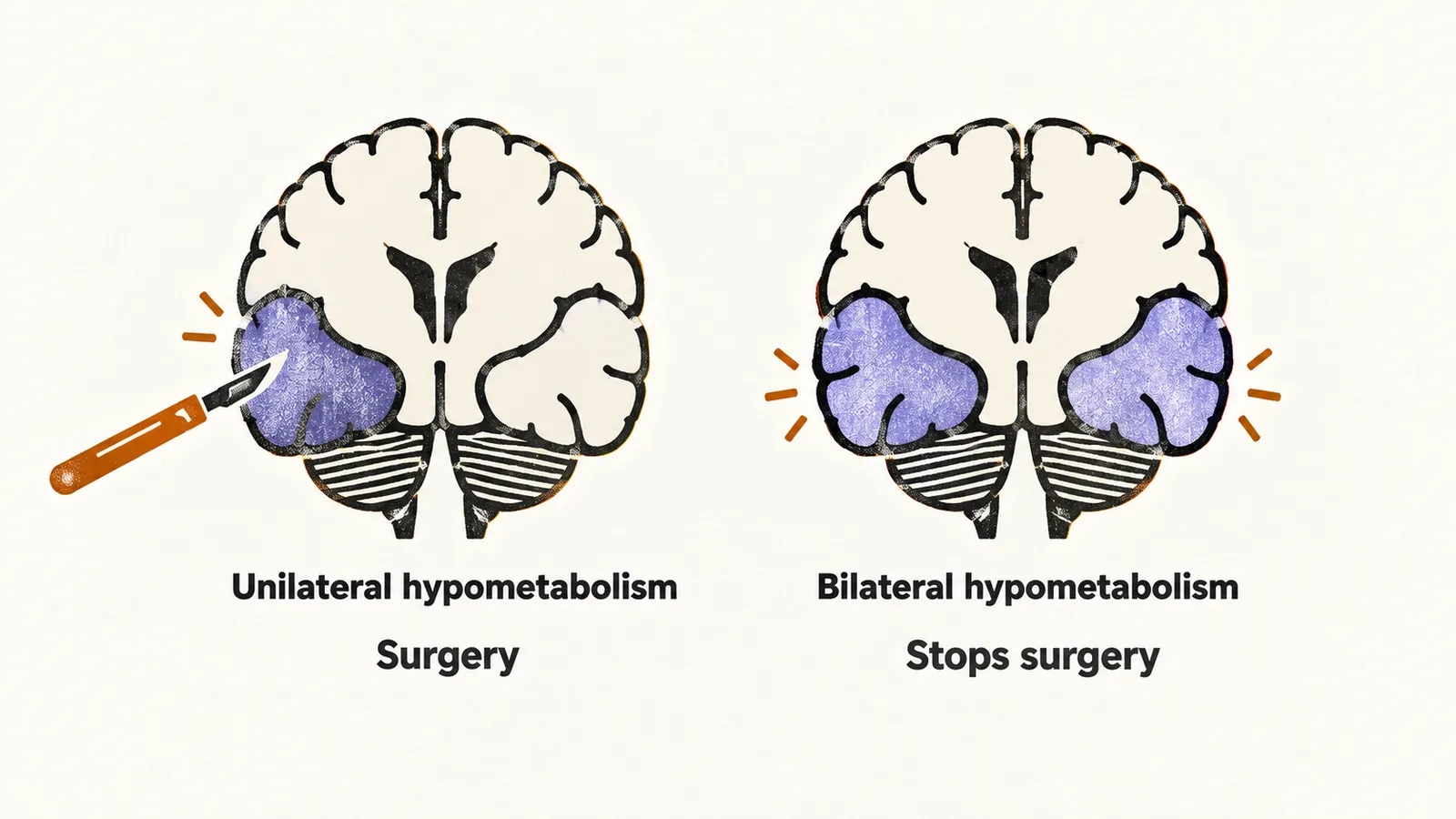
Temporal lobe epilepsy responds best to surgery, with reported cure rates around 90% for well-selected cases. FDG-PET lateralizes the focus as unilateral temporal hypometabolism, succeeding where serial EEG struggles: the two mesial temporal lobes are densely connected, so discharges appear on one side in one recording and the other in the next. Metabolism has no such mirror effect.
Correct lateralization is unforgiving, since only one temporal lobe can ever be removed; resecting both would destroy memory, and operating the wrong side leaves the seizures behind. Conversely, bilateral temporal hypometabolism stops surgery even when EEG favours one side, because the contralateral lobe would keep generating seizures. Preventing that futile operation is among PET’s most valuable contributions.
Outside the temporal lobe
Outside the temporal lobe, sensitivity falls. The extratemporal cortex is larger and more variable, asymmetries are subtler, and boundaries blur, so analysis grows harder. PET still detects focal hypometabolism that helps, especially when coregistered with MRI (the alignment of the PET image onto the anatomical scan), but expectations should be set lower than in the temporal lobe.