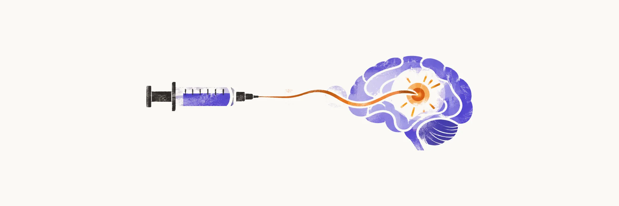
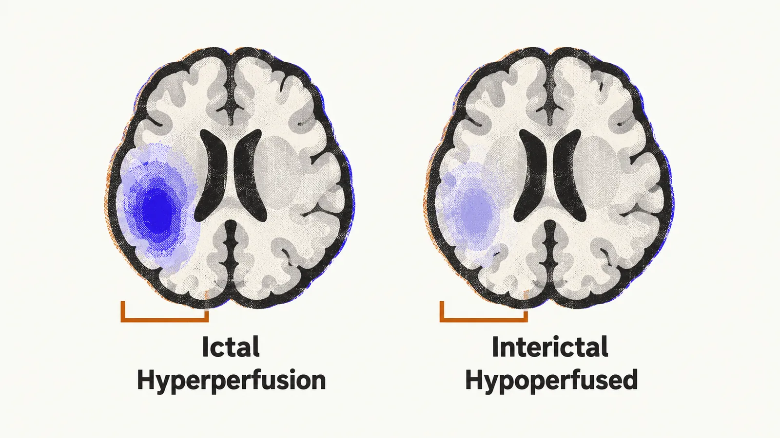
SPECT (single-photon emission computed tomography) uses a radioactive tracer that distributes through the brain in proportion to blood flow, and it exploits the perfusion flip between seizure states. During a seizure (the ictal state), the firing onset zone, the region where the seizure begins, demands more oxygen and glucose, driving cerebral blood flow up by at least half from a baseline near 50 mL per 100 g per minute. Between seizures (the interictal state) the same region runs hypoperfused, meaning it receives less blood flow than normal.
Because SPECT tracers distribute with blood flow at the instant of injection, an ictal scan marks the focus as a hot spot of hyperperfusion, meaning increased blood flow, while an interictal scan shows a cool zone. Comparing the two states localizes the onset zone.
Why timing decides the result
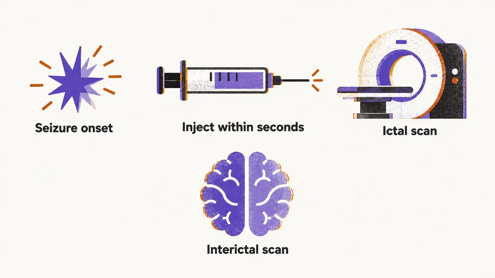
The technique images the seizure itself, which is its edge over FDG-PET (PET, positron emission tomography, with the glucose tracer fluorodeoxyglucose): PET accumulates signal over 30 to 45 minutes into a blended metabolic picture, while SPECT freezes one perfusion moment. That advantage costs logistics. The tracer must be injected within seconds of seizure onset, before activity propagates, so the patient waits in an epilepsy monitoring unit under continuous electroencephalography (EEG) with a prepared dose at the bedside, and a second interictal scan follows on a seizure-free day. Delays beyond roughly half a minute to a minute risk mapping propagated activity instead of the origin.
Resolution and complementing PET
SPECT resolution is coarser than PET, around 1 cm against 3 to 4 mm, yet when interictal PET is non-diagnostic or diffusely non-localizing, a focal ictal hyperperfusion can still supply the evidence surgery needs. The two techniques complement rather than compete.