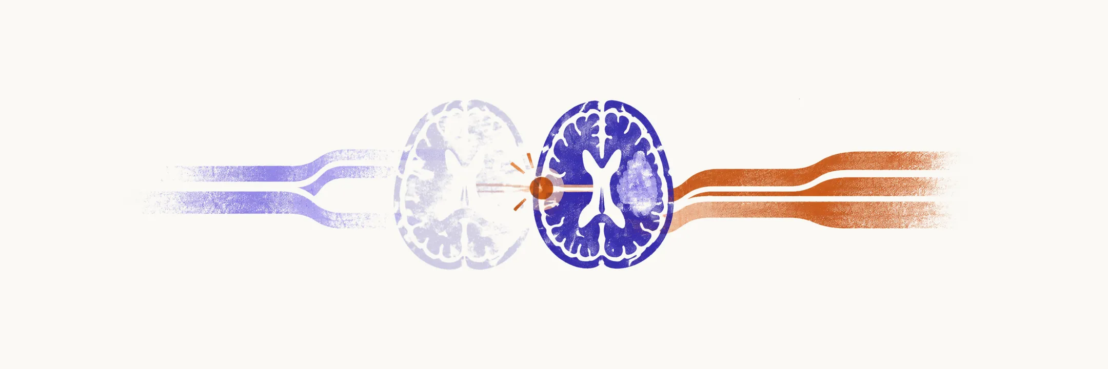
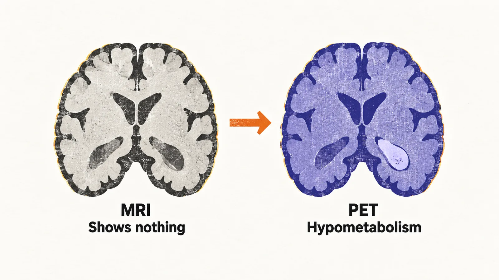
In drug-resistant epilepsy, about one-third of patients have a normal magnetic resonance imaging (MRI) scan, leaving the surgeon with an electroencephalography (EEG) suggestion of where seizures start and no visible target. FDG-PET, which images glucose metabolism between seizures, can reveal a region of reduced metabolism, or hypometabolism, where MRI shows nothing, including a hippocampus without signal change or atrophy, and thereby justify surgery that MRI alone would have foreclosed.
When MRI does show a lesion
A lesion on MRI does not make PET redundant. Take mesial temporal sclerosis, the hippocampal scarring seen in temporal lobe epilepsy: PET sizes the abnormality. If hypometabolism spans the whole hippocampus, a standard resection sparing its posterior part will likely leave epileptic tissue behind, and the surgeon must widen the resection.
PET can also show hypometabolism far from the lesion. Remote hypometabolism elsewhere in the brain tempers expectations symmetrically, suggesting secondary network damage or diffuse pathology that one resection will not cure.
Matching PET to anatomy
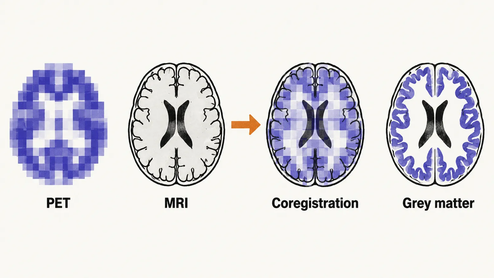
Either way, PET has to be read against anatomy. A high-quality MRI stays indispensable for reading PET: without anatomy, metabolic change cannot be assigned to lesion, normal cortex, or artefact. Simultaneous PET-MRI scanners are costly, and computationally registering a separately acquired PET volume onto high-resolution MRI (coregistration) achieves equivalent colocalization, since the brain holds its shape between scans. The rule is to register the lower-resolution PET onto the higher-resolution MRI and then inspect grey matter systematically.
Even a well-registered PET may not show a clear focus, and then a different signal, blood flow captured during a seizure itself, becomes the next thing to ask about.