
Because amenorrhea is a sign rather than a diagnosis, its evaluation starts with the cause behind it. Amenorrhea that is not caused by pregnancy, lactation or menopause affects approximately 3–4% of women of reproductive age.
Classification by cause
Amenorrhea is grouped by where the fault lies, and the three groups point to different parts of the axis:
- Anatomic amenorrhea: the uterus or the genital outflow tract is affected, so the endometrium cannot respond to hormones or the menstrual blood cannot leave the body.
- Ovarian failure: the ovary itself fails, producing little estrogen and few follicles; the loss of estrogen feedback is what raises the gonadotropins. Gonadotropins are the pituitary hormones follicle-stimulating hormone (FSH) and luteinizing hormone (LH), which drive the ovary.
- Chronic anovulation: the ovary is intact but ovulation is blocked by an endocrine disturbance elsewhere in the axis, in the hypothalamus or pituitary, or through abnormal steroid feedback.
| Category | Causes |
|---|---|
| Anatomic causes | Pregnancy, Müllerian agenesis or dysgenesis, cervical stenosis, disorders of sexual differentiation, (Asherman syndrome) |
| Ovarian failure | Menopause, genetic abnormalities (X chromosomal causes such as Turner syndrome and pure gonadal dysgenesis, and autosomal causes), immune dysfunction, physical insults (chemotherapy, radiation), idiopathic |
| Chronic anovulation | Hypothalamic causes (psychogenic, exercise-associated, eating disorders, systemic illness, neoplasms), pituitary causes (pituitary tumours and hyperprolactinemia), inappropriate steroid feedback (polycystic ovary syndrome or PCOS, adrenal hyperplasia, various neoplasms), other endocrine disorders (thyroid dysfunction, adrenal hyperfunction) |
History and physical examination
The three categories can often be told apart at the bedside, because each leaves different signs. In most patients the history and physical examination suggest the diagnosis before any test is ordered, so this is the most important step. Four aspects are examined because each points toward a different group of causes:
- body dimension and habitus — weight loss, low body weight or obesity point toward functional hypothalamic causes or PCOS;
- distribution and presence of androgen-stimulated body hair — hirsutism (excess androgen-stimulated body hair) points to androgen excess, most often PCOS;
- extent of breast development and presence or absence of breast secretions — breast development shows previous estrogen action, meaning the ovary has worked before, whereas galactorrhea (milk secretion from the breast) points to hyperprolactinemia, an elevated prolactin level;
- external and internal genitalia — an absent or blind vagina, or an absent uterus, points to an anatomic cause.
Excluding pregnancy
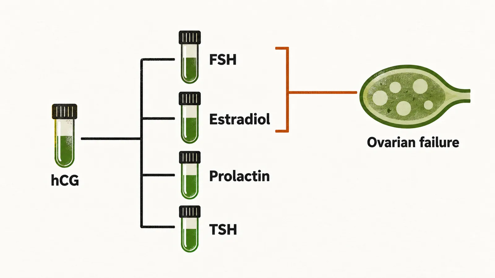
The most common cause of secondary amenorrhea is pregnancy, so pregnancy is evaluated first with a human chorionic gonadotropin (hCG) test. A urinary test can occasionally be falsely negative, so a serum hCG is preferred when clinical suspicion is high.
First-line laboratory tests
The initial workup then consists of four tests, FSH, estradiol, prolactin and thyroid-stimulating hormone (TSH):
- FSH level and estradiol (E2) level are interpreted together, because they separate ovarian failure from the other causes. In ovarian failure the ovary no longer produces estrogen, so E2 is low while FSH is high; in most other causes FSH is normal or low. An elevated FSH in a woman under 40 points toward .
- Prolactin level: hyperprolactinemia suppresses the gonadotropin signal and is a common, treatable cause of amenorrhea, but only about one third of women with hyperprolactinemia have galactorrhea, so its absence does not exclude the diagnosis. A mildly elevated level (20–40 ng/mL) should be repeated before the diagnosis is made.
- TSH level: both hypothyroidism and hyperthyroidism disturb ovulation, and treating the thyroid disorder usually restores spontaneous menses.
A was traditionally used to separate anovulation from other causes: withdrawal bleeding means the endometrium has been estrogenised and the problem is anovulation, while no bleeding means either low estrogen or an outflow-tract problem. Because measuring FSH and E2 now answers the same question more directly, the challenge is less often needed. A high FSH with a low E2 points to ovarian failure, and the next question is what has caused it.