Hypergonadotropic amenorrhea is amenorrhea in which the gonadotropins — the pituitary hormones FSH and LH that drive the ovary — are high. The high level is the pituitary’s response to an ovary that has stopped producing estrogen and follicles: without the negative feedback of ovarian estrogen, gonadotropin secretion rises.
Primary ovarian insufficiency
One of the main causes of hypergonadotropic amenorrhea is primary ovarian insufficiency or POI, in which there is a triad of:
- amenorrhea
- hypergonadotropinism
- hypoestrogenism
The diagnosis is made when menstrual cycles are disordered (spontaneous amenorrhea or irregular cycles) for at least 4 months with an FSH level above 25 IU/L in a woman under 40 years of age. FSH does not have to be measured on a particular day of the cycle, but a raised value should be repeated if there is diagnostic uncertainty, and pregnancy should be excluded first. POI can present as either primary or secondary amenorrhea.
Primary ovarian insufficiency is a very heterogeneous condition, and its causes range from a single genetic mutation, cytological chromosomal abnormalities and a single enzymatic mutation or deficiency to environmental causes. Genetic causes are the most important single group, although a specific genetic cause is identified in only a minority of cases; the premutation is the most common single-gene cause, found in about 2% of women with sporadic POI and about 14% of those with a familial form.
Terminology of gonadal dysgenesis
Several causes of POI involve gonadal dysgenesis, in which the primary gonad — the ovary or the testis — does not develop, or is under-developed. It comes in three forms:
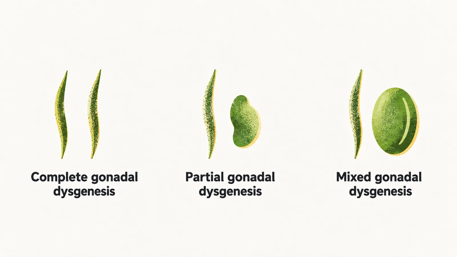
- Complete gonadal dysgenesis: the gonads are entirely replaced by streak gonad, a fibrous streak of tissue.
- Partial gonadal dysgenesis: there is some degree of gonadal development, but it is not complete, and it can lead to ambiguous genitalia.
- Mixed gonadal dysgenesis: it happens due to genetic mosaicism, the presence of more than one cell line with a different chromosome complement in the same person, which is characterised by the presence of one streak gonad and one partially developed gonad on the other side.
Causes of POI
POI can be caused by genetic, autoimmune and environmental causes. The genetic group is the most important and comes first: disorders of gonadal development, changes in single genes such as FMR1, the enzymes of steroid synthesis and the FSH receptor.
Genetic causes
Gonadal dysgenesis. An intact 46,XX karyotype (the chromosome complement of a typical woman) is necessary for the ovary to develop and for oocytes to survive, so depletion or mutation of X-chromosomal material can exhaust the ovary and lead to POI. Three X-chromosome loci are named for this association — POF1 (Xq26–q28, which includes the FMR1 gene), POF2 (Xq13.3–q21.1) and POF4 (Xp11.2, the BMP15 gene) — and mutations at these loci are among the identified causes. Gonadal dysgenesis can be complete, partial or mixed.
Complete gonadal dysgenesis happens in women in two situations.
In 46,XX dysgenesis, women with 46,XX genetics who have apparent female characteristics, a uterus and a vagina, but an infantile form with streak gonads, carry mutations needed for the development of the ovaries; these mutations can be either autosomal or X-linked and leave the ovaries as fibrous streaks.
In 46,XY dysgenesis, subjects are genetically male, but they have a mutation in the SRY gene (or in another gene needed for testis determination), so the testes do not develop. Without testes there is no anti-Müllerian hormone and no testosterone, so the internal and external genitalia are female; the gonads remain streaks and the person is infertile.
Complete gonadal dysgenesis with streak ovaries must be told apart from Turner syndrome. In Turner syndrome there are characteristic physical findings, but in pure gonadal dysgenesis there are no specific findings of Turner syndrome: subjects have normal height and weight, normal genitalia, but streak ovaries.
Partial gonadal dysgenesis leaves some degree of gonadal development.
Mixed gonadal dysgenesis is most often caused by mosaicism, and most patients carry the genetics of 45,X/46,XY, so there is one streak gonad and one partially developed testis. The vast majority of the cases are raised as females. Because the dysgenetic gonad carries a high malignant potential, it should be removed surgically.
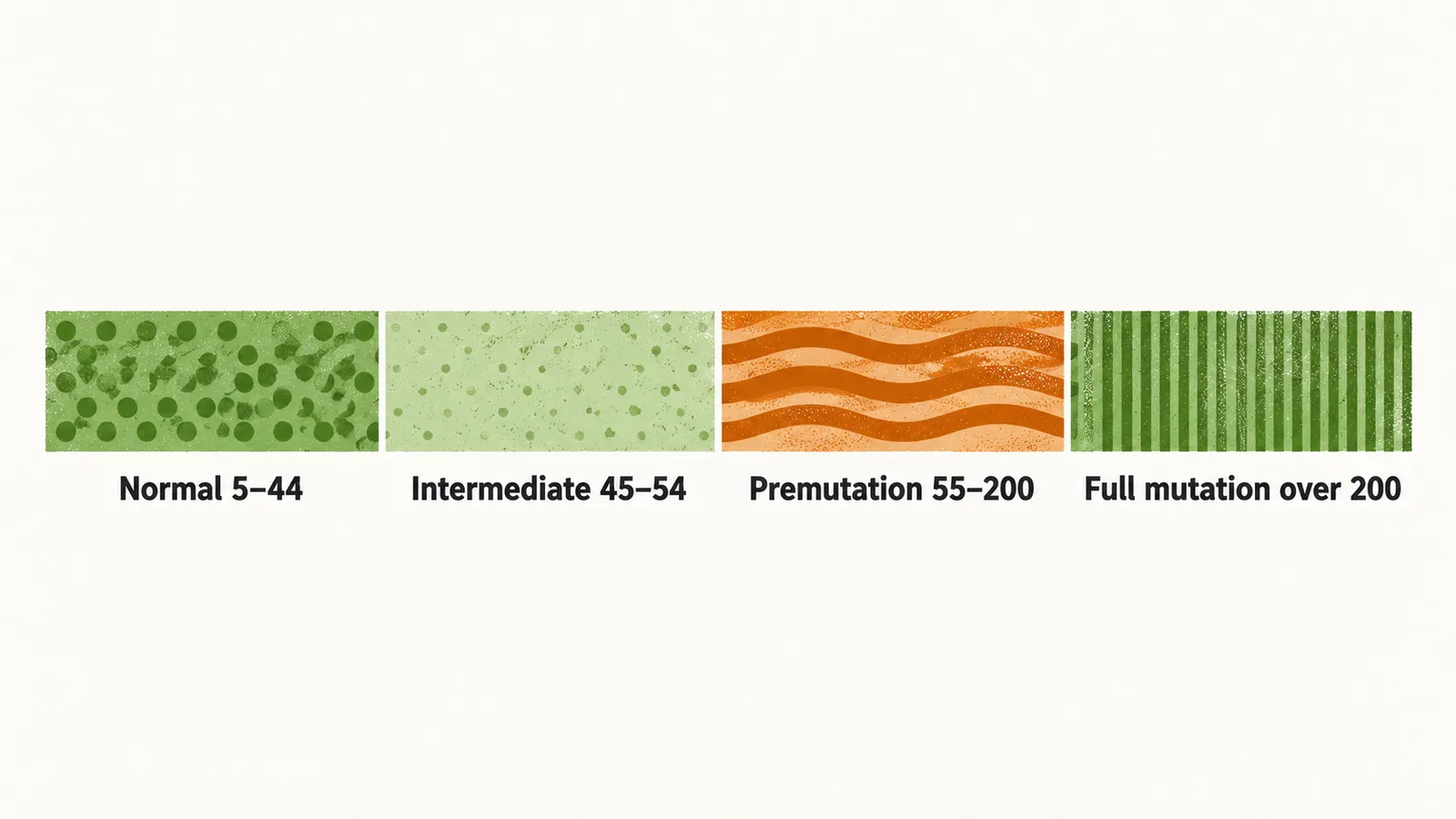
FMR1 premutation. The FMR1 gene lies on the X chromosome and normally has 5–44 CGG repeats. Longer expansions are classified as intermediate (45–54 repeats), premutation (55–200 repeats) and full mutation (more than 200 repeats). In the premutation state the gene still produces mRNA, but the expanded mRNA is toxic to the ovarian cells and accelerates follicular atresia, so carriers are at risk of POI; the premutation is the most frequent single-gene cause. In the full mutation state the gene is silenced and its function is lost, which causes fragile X syndrome: the presentation is prominent in males but milder in females, because one X chromosome is largely inactivated. A woman with a premutation can pass on more repeats, because expansion occurs during maternal transmission, while expansion does not occur when a premutation is passed by a father.
Enzymatic mutations. A (a mutation of CYP17A1) in girls who survive to the teenage years blocks cortisol and sex steroid synthesis and can lead to POI, with an increased progesterone level, increased FSH and LH, and increased deoxycorticosterone and corticosterone; the mineralocorticoid excess causes hypertension with a low potassium level, while aldosterone itself is suppressed. 17,20-desmolase and 20,22-desmolase mutations affect the same steroid pathways. An aromatase mutation prevents estrogen synthesis: these patients are manifested at birth by clitoromegaly and posterior labioscrotal fusion, have severe estrogen deficiency, and are virilized by the high androstenedione level that cannot be converted to estrogen.
FSH resistance. An FSH receptor mutation — the gene lies on chromosome 2 — can lead to failure of folliculogenesis, and this condition is fairly rare in many populations.
Other genetic mutations include the inhibin alpha gene (INHA), the BMP15 gene and the AIRE gene.
Autoimmune causes
POI can be associated with other endocrinopathies. The most common autoimmune association in women is thyroiditis, and POI can also be seen in patients with poly-endocrine autoimmune conditions that present with hypoparathyroidism, hypoadrenalism and mucocutaneous candidiasis. In a restricted ovarian autoimmune condition the immune attack is confined to the ovary.
Environmental causes
The ovary can also be damaged directly, by ionizing radiation, by chemotherapeutic agents or by viral infections.