Cushing’s syndrome, the state of chronic glucocorticoid excess, is easy to miss when the signs are mild, because obesity, depression, diabetes, hypertension, and menstrual irregularity are all common in the general population. The diagnosis is therefore made in two stages: first confirm that cortisol excess is present, then find its cause. A morning cortisol level on its own is not a good screening test.
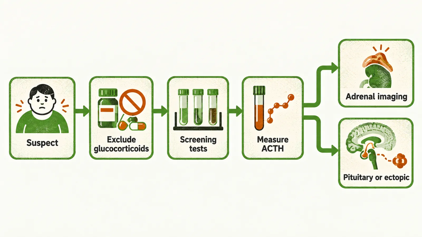
When to suspect Cushing’s syndrome
Cushing’s syndrome should be considered when:
- a patient has features of the syndrome that cannot be explained in another way;
- several features cluster in a pattern that does not fit a single diagnosis;
- the features appear at a much earlier age than expected (for example osteoporosis, fractures, or hypertension in a young adult); or
- the features accumulate over a period of time.
It should also be considered as a secondary cause in patients with obesity, depression, diabetes, hypertension, or menstrual irregularity who have additional features that those conditions do not explain, such as wide purple striae or proximal muscle weakness.
Excluding exogenous glucocorticoids
Exogenous glucocorticoids, meaning glucocorticoids taken as medication, are the most common cause of Cushing’s syndrome and are easy to overlook, because they are prescribed by several routes — oral, topical, rectal, and inhaled — and are present in some injections, non-prescription topical medicines, skin-lightening creams, and traditional herbal preparations. A Cushingoid patient (one with the features of Cushing’s syndrome) who may have used any of these is assessed for exogenous exposure before an endogenous cause is pursued.
Screening tests
Initial screening uses a test with high diagnostic accuracy: 24-hour urinary free cortisol (UFC), late-night salivary cortisol, or the 1-mg overnight dexamethasone suppression test, with the 2-mg 2-day dexamethasone suppression test as an alternative. Dexamethasone is a synthetic glucocorticoid that normally suppresses cortisol through negative feedback. The tests reflect different abnormalities — high integrated daily cortisol production for UFC, loss of the late-night cortisol nadir (the normal low point) for salivary cortisol, and impaired negative feedback for dexamethasone suppression — so they complement each other and more than one is usually measured. Because cortisol secretion varies over time, at least two measurements are needed for UFC and for late-night salivary cortisol.
After 1 mg of dexamethasone is taken at midnight, a normal cortisol at 9 a.m. is below 1.8 µg/dL (50 nmol/L), and higher values indicate autonomous secretion. The result depends on the assay used, so it must be interpreted against that assay’s own reference range.
Mildly abnormal results also occur in conditions that raise cortisol without true Cushing’s syndrome, such as pseudo-Cushing’s states driven by stress, depression, or anorexia nervosa (see Causes and Forms of Cushing’s Syndrome); these are usually separated by observing the patient over time and treating the underlying condition.
Finding the cause
Once cortisol excess is confirmed, ACTH (adrenocorticotropic hormone, the pituitary hormone that drives the adrenal cortex) is measured as the first step. A low or undetectable ACTH identifies an ACTH-independent cause, and the adrenal glands are then imaged with CT or MRI to look for a unilateral mass with atrophy of the opposite gland, or for bilateral disease. A normal or elevated ACTH identifies an ACTH-dependent cause, which is investigated further to separate a pituitary from an ectopic source using a high-dose (8 mg) dexamethasone suppression test, a corticotropin-releasing hormone (CRH) stimulation test, pituitary MRI, and inferior petrosal sinus sampling, in which blood draining the pituitary is sampled.
Localising the source in ACTH-dependent disease
In Cushing’s disease the adenoma is usually a microadenoma and is visible on pituitary MRI in only about 60% of adults. A pituitary lesion is not proof of Cushing’s disease, because about 10% of healthy adults have small pituitary lesions visible on MRI, and about 12% of patients with ectopic ACTH secretion also have an abnormal pituitary MRI. When imaging is negative or ambiguous, inferior petrosal sinus sampling is used to distinguish a pituitary from a non-pituitary source; it does not reliably predict the side of the tumour. For a suspected ectopic source, imaging of the chest and abdomen is guided by the likely tumour.