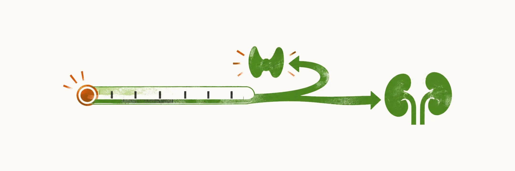
Diagnosing hypocalcemia means confirming that the calcium is genuinely low and then identifying which cause produced it. The two steps are linked, because a low total calcium is not always a low ionised calcium, and because the same low calcium means different things depending on whether the parathyroid glands are answering.
Confirming that the calcium is low
The first step is to confirm the result and to rule out an artefactually low calcium. About 40% of the calcium in serum is bound to albumin (with a further fraction bound to other anions), so a low total calcium in a patient with hypoalbuminemia (a low serum albumin) may not represent a low ionised calcium, the fraction the tissues respond to. In that situation the ionised calcium can be measured directly, or the total calcium can be corrected for the albumin:
In critically ill patients with acid-base disturbances, ionised calcium is preferable, because the binding of calcium to albumin changes with pH. An ionised calcium below about 1.17 mmol/L (4.7 mg/dL) is low.
Finding the cause
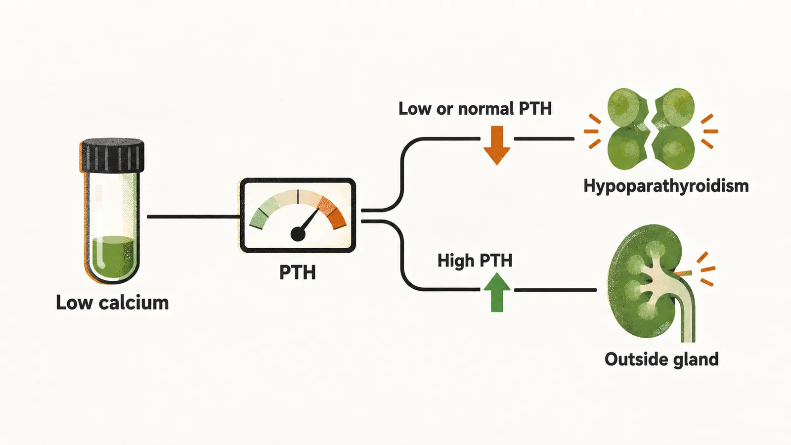
Serum parathyroid hormone (PTH) is the most useful test, and it is read in the context of the calcium level rather than on its own. When the calcium is low, PTH should be high. A low, or inappropriately normal, intact PTH means the glands are not responding, which is hypoparathyroidism. A high PTH means the glands are responding, so the fault lies outside them, in a cause such as vitamin D deficiency, chronic kidney disease, malabsorption, or PTH resistance. The assay used should be an intact PTH assay with reliable performance at the low end of the normal range.
Measuring serum phosphate, creatinine and 25-hydroxyvitamin D alongside PTH usually identifies the cause. Phosphate tends to be high when PTH is absent or ineffective, and low when PTH is high, so it helps separate hypoparathyroidism and pseudohypoparathyroidism from vitamin D deficiency, in which the rise in PTH keeps phosphate low. Magnesium should always be checked, because magnesium depletion both impairs the secretion of PTH and blunts its action on the kidney; hypocalcemia caused by magnesium deficiency does not correct adequately with calcium and vitamin D until the magnesium is replaced. Further tests are added according to the presentation, and can include lipase for pancreatitis, ACTH and cortisol and TSH for polyendocrine failure, and 1,25-dihydroxyvitamin D for deficiency states.
Recognising pseudohypoparathyroidism
A patient who presents with the signs and symptoms of hypocalcemia while the serum PTH is high should raise the suspicion of pseudohypoparathyroidism.
Albright hereditary osteodystrophy, or AHO, is typically manifested as:
- short stature
- brachydactyly
- subcutaneous calcification
- obesity
In a patient who presents with AHO, the diagnosis to consider is either pseudohypoparathyroidism 1a or pseudopseudohypoparathyroidism. A patient with only AHO and no biochemical alteration related to resistance to PTH has PPHP, with a normal serum calcium and a normal serum phosphate.
The classical test for pseudohypoparathyroidism measures the response of the kidney to an injection of PTH, through the excretion of cyclic AMP (cAMP) in the urine and the level of phosphate in the urine. A normal physiological response, and the response in hypoparathyroidism, is a 10- to 20-fold increase of urinary cAMP, whereas in PHP 1a and PHP 1b there is no increase. The definitive diagnosis of PHP 1 is genetic analysis of the GNAS gene.
Once the cause is identified, the next question is how far the calcium has fallen and whether it is causing symptoms, because that decides how quickly it must be replaced.