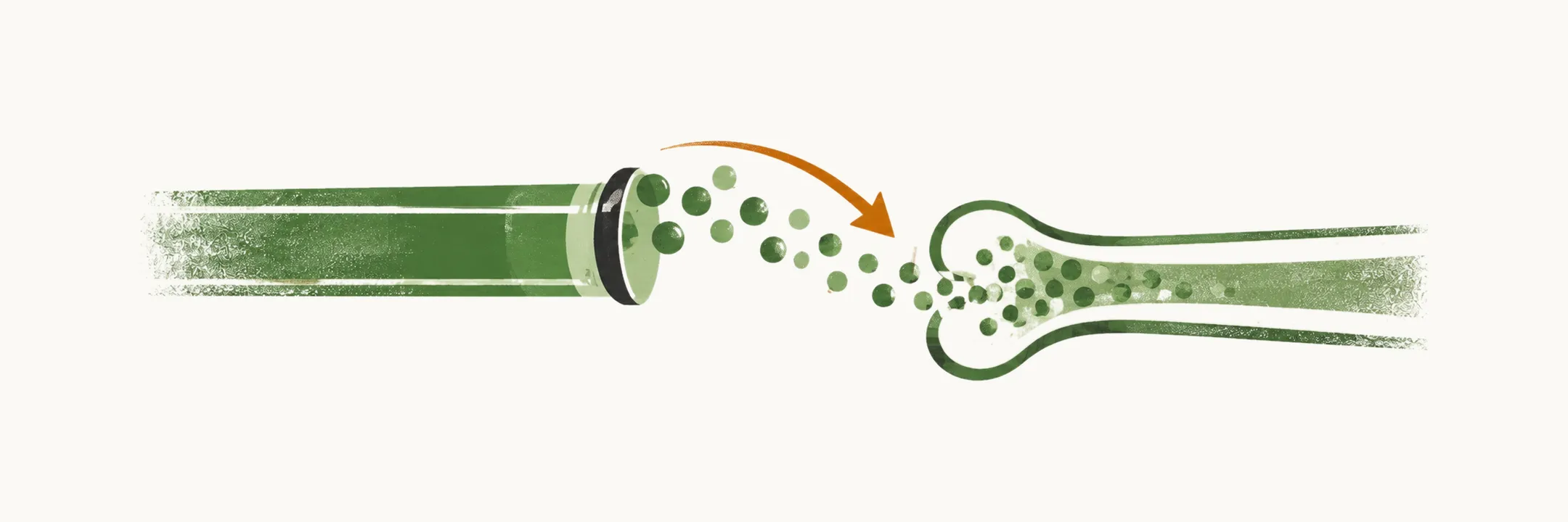
Hypocalcemia has a limited set of causes, and they can be grouped by which part of calcium control has failed: the vitamin D pathway, the parathyroid response (the release of parathyroid hormone, PTH), or the traffic of calcium out of the blood and into bone or damaged tissue. The mechanism behind each cause explains the calcium level that results and points to the treatment it needs.
Vitamin D deficiency
Acquired or inherited vitamin D disease is one of the most common causes of hypocalcemia. The estimated prevalence of true vitamin D deficiency, defined as a 25-hydroxyvitamin D level below 20 ng/ml, is about 40%.
The acquired causes are a low intake of food fortified with vitamin D or a restricted diet, cultures in which clothes cover most of the body, malabsorption, and liver and renal disease. Liver disease is not a common cause of vitamin D deficiency, because at least 90% damage to the liver is needed to affect the production of 25-hydroxyvitamin D directly; in the early stages of liver disease, however, intestinal vitamin D absorption is abnormal, because fat absorption is abnormal. Renal disease lowers vitamin D in two different ways: in nephrotic syndrome vitamin D falls, possibly because the carrier proteins for vitamin D are lost from the serum, while in chronic renal failure hyperphosphatemia reduces the production and activation of vitamin D.
The inherited causes are rickets and the vitamin D-dependent rickets (VDDR) disorders. Rickets is a spectrum of disorders in which growing bone is not mineralised normally, so the bones stay soft and deform; it can be caused by deficiency of vitamin D, calcium or phosphate. Vitamin D-dependent rickets is very uncommon and occurs in two forms: type 1 results from a defect in the renal production of 1,25-dihydroxyvitamin D, and type 2 from a mutation in the vitamin D receptor.
Hypoparathyroidism
Hypoparathyroidism means inadequate PTH production, and its most common cause is post-surgical damage, which can happen during surgery on the parathyroid or the thyroid gland. Permanent post-operative hypoparathyroidism is diagnosed when the hypoparathyroidism is still present at least 6 months after surgery; in some cases, therefore, it resolves spontaneously. Autoimmune hypoparathyroidism is the second most common cause of hypoparathyroidism in adults.
Post-operative hypocalcemia
Transient hypocalcemia after thyroidectomy is the pattern encountered most often. The proportion of patients affected differs between studies; in one study the figure is estimated at around 35%, with the permanent form in about 2% of cases. Some studies suggest a cutoff for predicting symptomatic hypocalcemia after total or near-total thyroidectomy. The test is the 1-hour post-operative intact PTH: a low level means a higher chance of hypocalcemia, and an intact PTH above 10 pg/ml means these patients do not develop symptomatic hypocalcemia.
Hypocalcemia after parathyroidectomy for an adenoma follows a different route: the remaining, normal parathyroid glands were suppressed by the adenomatous gland that has been removed, and they need time to regain their function.
Genetic syndromes that cause hypoparathyroidism
Hypoparathyroidism can also be one part of a genetic syndrome. Two are described here.
DiGeorge syndrome
DiGeorge syndrome is the most common gene deletion syndrome in humans, occurring in 1 in every 4000 newborns. A microdeletion of part of chromosome 22 disrupts the development of the third and fourth pharyngeal pouches, and the result is aplasia or hypoplasia of the thymus gland, so immunodeficiency and recurrent respiratory infections are common; aplasia or hypoplasia of the parathyroid glands; and cardiac outflow malformations.
Autoimmune polyendocrine syndrome type 1
Hypoparathyroidism can also occur in an autoimmune context, autoimmune polyendocrine syndrome type 1 (APS-1), which is caused by a mutation in the AIRE gene. At least two of three conditions are needed for the diagnosis:
- autoimmune parathyroid disease
- autoimmune adrenal disease
- mucocutaneous candidiasis
The classic triad is hypoparathyroidism, candidiasis and adrenal insufficiency (Addison’s disease). Two or three of the following are also associated: type 1 diabetes mellitus, primary hypogonadism (especially ovarian failure), autoimmune thyroid disease, chronic active hepatitis, alopecia (totalis or areata), and vitiligo.
Hungry bone syndrome
Hypocalcemia after thyroidectomy or parathyroidectomy does not always come from a failing parathyroid gland. After either operation in a patient with hyperparathyroidism, bone can mineralise rapidly, a state called hungry bone syndrome. It is thought to happen because the sudden loss of the stimulus to bone resorption, which could be PTH or thyroid hormone, leaves bone formation running ahead of resorption, so mineral is drawn out of the blood after the operation. The rapid mineralisation of bone causes bone pain, and the resulting hypocalcemia causes tetany.
Other causes
Several further causes do not belong to the groups above. The miscellaneous etiologies include neonatal hypocalcemia and hyperphosphatemia, which arises from phosphate retention in acute or chronic renal failure, excess phosphate absorption caused by enemas or oral supplements, or massive phosphate release caused by tumour lysis or crush injury. Drugs can also cause hypocalcemia, particularly intravenous bisphosphonate or denosumab therapy in patients with vitamin D insufficiency or deficiency, and foscarnet. The remaining miscellaneous etiologies are rapid transfusion of large volumes of citrate-containing blood; acute critical illness; hungry bone syndrome after thyroidectomy for Graves’ disease or after parathyroidectomy; osteoblastic metastases; acute pancreatitis; rhabdomyolysis; and mitochondrial gene defects.
All the causes above, grouped by the failure they represent, are:
- Inadequate vitamin D production and action: nutritional deficiency; lack of sunlight exposure; malabsorption; post-gastric bypass surgery; end-stage liver disease and cirrhosis; chronic kidney disease; vitamin D-dependent rickets types 1 and 2.
- Inadequate PTH production (hypoparathyroidism).
- Functional hypoparathyroidism: magnesium depletion and magnesium excess.
- PTH resistance: pseudohypoparathyroidism, in which the parathyroid gland makes PTH but the tissues do not answer it.
- Miscellaneous etiologies: as listed above.