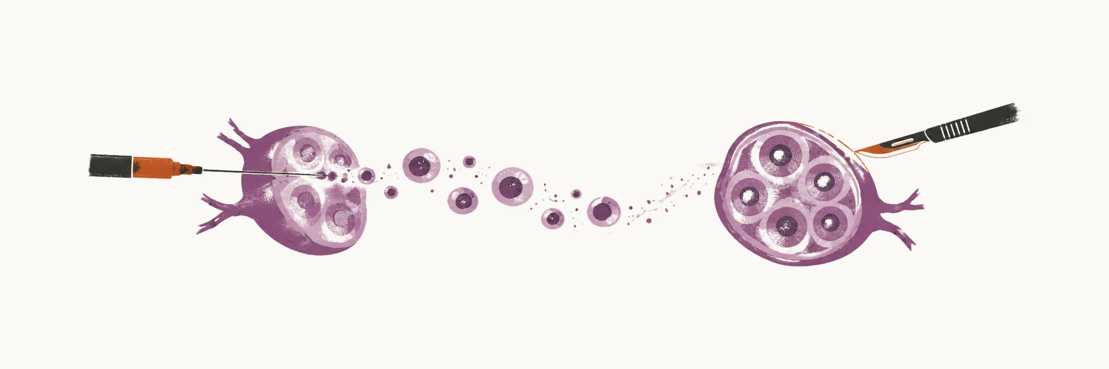
When non-Hodgkin lymphoma (NHL) is suspected, the diagnosis is confirmed on tissue, and the choice of test is driven by what the diagnosis requires: an intact piece of tumour, not only a sample of its cells.
Tissue biopsy
The diagnosis rests on a tissue biopsy — usually excisional (removing a whole node) or tru-cut (core needle) — because the subtype is defined by the tumour’s architecture and its marker pattern, and both are lost when a needle removes only loose cells. A fine needle alone is therefore not sufficient. Alongside the biopsy, the workup looks for disease in the sites it likes to reach:
- bone marrow biopsy — detects marrow involvement, which is common in the indolent subtypes
- lumbar puncture — detects central nervous system (CNS) involvement
- laboratory tests — complete blood count (CBC), virology and serum electrophoresis
- imaging — CT and PET-CT; PET-CT adds a map of metabolic activity to the anatomical detail of CT, so it shows which nodes and extranodal sites are actually involved
Staging
The disease is then staged by how many nodal and extranodal sites (sites outside the lymph nodes) are involved and where they lie, which is why the imaging and marrow studies are done together. The stage and the sites of involvement are two of the factors that set the prognosis and the treatment.