
The different subtypes of non-Hodgkin lymphoma (NHL) are largely set apart by the cytogenetic abnormalities they carry, meaning the chromosomal changes found in the tumour cells, and those same abnormalities explain which markers appear on the cells.
How translocations activate oncogenes
A translocation is the exchange of segments between chromosomes. Many lymphoid neoplasms carry a translocation that moves a proto-oncogene (a normal growth-promoting gene) next to the strong regulatory elements of an immunoglobulin (Ig) gene in B cells, or of a T-cell receptor (TCR) gene in T cells. Those regulatory elements then switch the oncogene on permanently, and the cell proliferates under a signal it cannot turn off. The Ig genes lie on chromosomes 2, 14 and 22; the TCR genes lie on chromosomes 7 and 14.
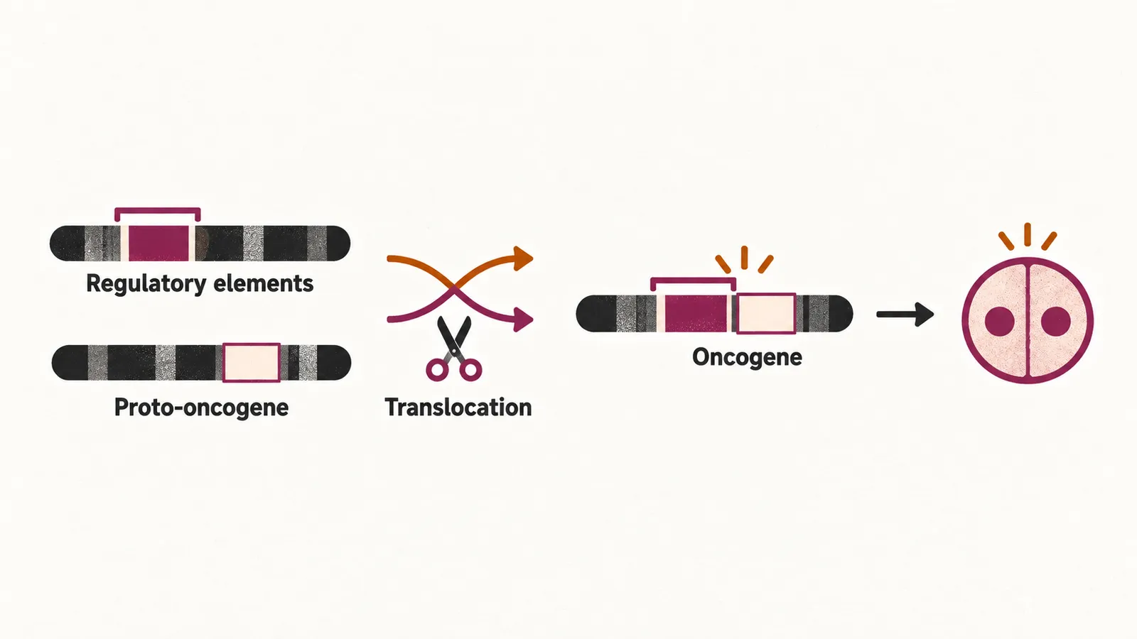
Recurrent translocations and transcription factors
Some of these changes involve transcription factors, proteins that switch other genes on or off. In B-cell lymphoma the recurring translocations involve the Ig chain genes on chromosomes 2, 14 and 22, and each pairs a specific oncogene with the Ig locus:
| Translocation | Lymphoma |
|---|---|
| t(8;14) | Burkitt lymphoma |
| t(11;14) | mantle cell lymphoma |
| t(14;18) | follicular lymphoma |
B-cell lymphomas also commonly carry abnormalities of MYC, BCL6 and BCL2. A separate transcription factor, PAX5 (also called BSAP), maintains B-cell identity and regulates Ig production, and it is expressed in nearly all B-cell lymphomas, where it serves as a marker of B-cell lineage.
In T-cell lymphoma the most important transcription factor is NOTCH1, and the recurring translocations involve the TCR genes on chromosomes 7 and 14.
Mutations of pathological significance
Beyond the translocations, point mutations (changes within a single gene) can carry pathological weight. TP53 mutation disables the tumour-suppressor response to DNA damage, and MYD88 mutation produces constitutive signalling (signalling that stays on without an external trigger) through the NF-κB pathway.
Identifying the markers of each level of differentiation
NHL is the neoplasm of mature B, T and NK cells, so its subtypes are distinguished by the proteins the cells express at each level of differentiation. Flow cytometry (which detects these proteins on cells in suspension) and immunohistochemistry (which detects them on tissue sections) are the methods used to find these markers on the tumour cells. Because a translocation usually leaves a characteristic marker pattern behind, the genetic and immunophenotypic findings are read together to assign the subtype.