
MS produces discrete focal neurological episodes, called relapses or attacks, whose symptoms depend entirely on where the lesions sit. Onset typically unfolds over days to weeks rather than minutes, which helps separate demyelination from vascular events. The most common presenting symptoms are weakness and sensory loss, followed by diplopia (double vision), vertigo, and urinary problems.
Clinical phenotypes
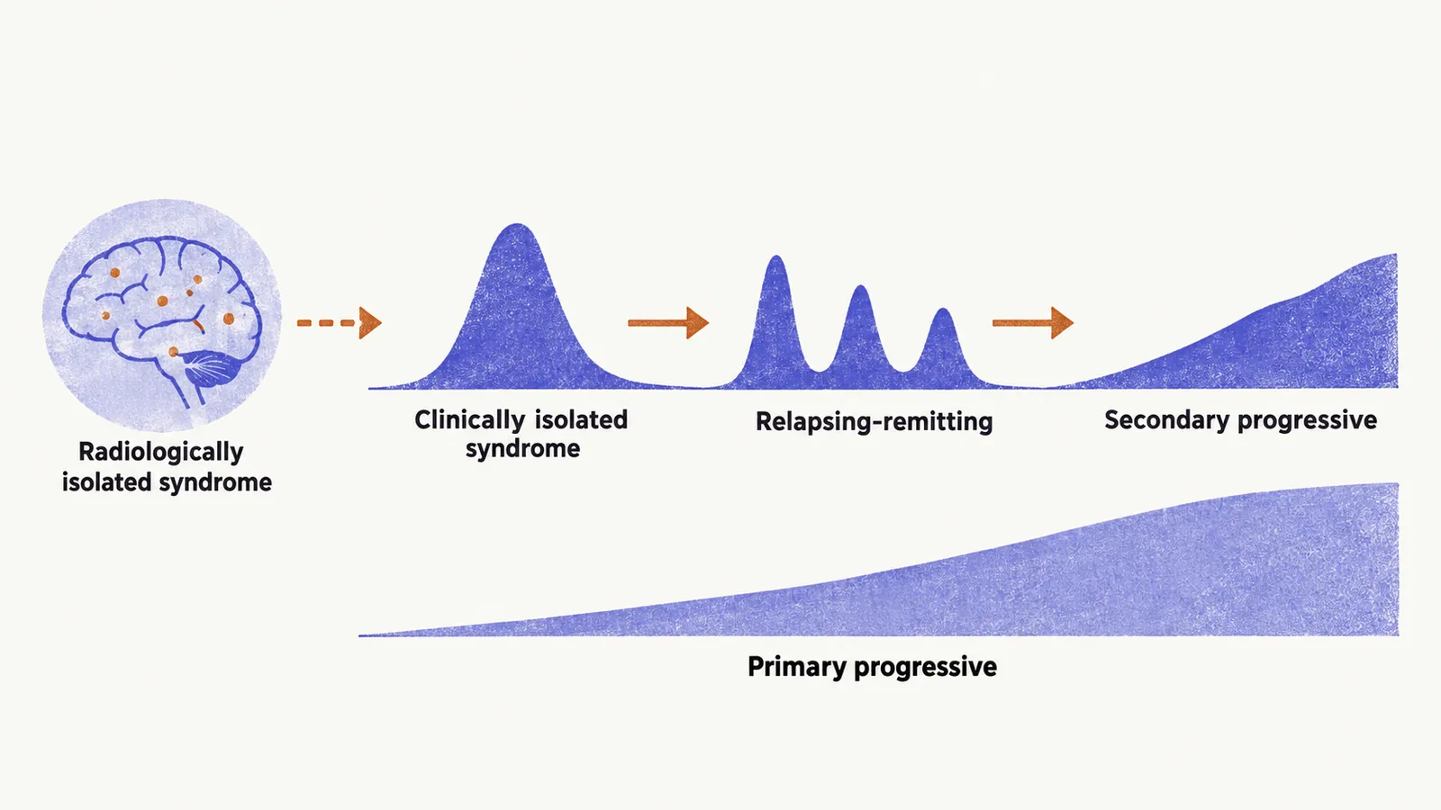
The phenotypes describe how the disease behaves over time: whether attacks come and go, whether disability accumulates on its own, and whether the disease is visible only on imaging.
Clinically isolated syndrome (CIS) is the first clinical episode suggestive of inflammatory demyelination, lasting more than 24 hours, that has not yet shown dissemination in time. When MRI already shows compatible lesions, the risk of developing clinically definite MS on follow-up is about 80%.
Relapsing-remitting MS (RRMS) accounts for roughly 85% of cases at onset. The disease alternates between discrete attacks and periods of stability. Historically about half of RRMS cases converted to secondary progressive disease within 10 years, and about 90% within 25 to 30 years. With modern disease-modifying therapy the reported conversion rate is roughly 18% after about 17 years.
Secondary progressive MS (SPMS) develops from RRMS when disability accumulates independently of relapses. Roughly 65% of RRMS cases were historically described as eventually transitioning. Two descriptors now refine the picture across phenotypes: relapse-associated worsening versus progression independent of relapse activity, which is disability accrual without recent relapses.
Primary progressive MS (PPMS) accounts for roughly 10 to 15% of cases, with progressive disability from onset and no initial relapses. Diagnosis requires at least 1 year of progression independent of relapses plus supporting evidence from brain MRI, spinal cord MRI, or cerebrospinal fluid.
Radiologically isolated syndrome (RIS) describes MRI findings suggestive of MS in a person without typical symptoms and with a normal examination. About two-thirds show radiological progression on later scans, and roughly one-third develop a clinical event within 2 to 5 years. Predictors of conversion include cervical cord or infratentorial lesions, higher lesion load, abnormal visual evoked potentials, younger age, positive cerebrospinal fluid, and more than 9 T2 lesions.
Disease activity is further described as active (a clinical relapse or MRI activity such as a gadolinium-enhancing or new or enlarging T2 lesion) or not active, and as progressive (worsening disability measured at least yearly, independent of relapse) or not progressive. These modifiers guide treatment decisions.
Typical features
Typical presentations follow the sites where plaques commonly sit. In the visual and brainstem pathways, they include acute unilateral optic neuritis (inflammation of the optic nerve), diplopia from bilateral internuclear ophthalmoplegia (impaired eye adduction from a lesion of the medial longitudinal fasciculus) or sixth-nerve palsy, facial numbness or trigeminal neuralgia, and cerebellar ataxia with nystagmus. In the spinal cord, they include partial myelopathy (a cord syndrome) with sensory loss and the Lhermitte sign, an electric-shock sensation down the spine on neck flexion from demyelinated cervical axons. Other typical features are asymmetric weakness, urinary incontinence, and fatigue.
Bilateral internuclear ophthalmoplegia is particularly suggestive because a midline lesion of the medial longitudinal fasciculus on both sides implies a central demyelinating process rather than a unilateral vascular one.
Atypical features
Some presentations should redirect thinking toward mimics such as neuromyelitis optica spectrum disorder, myelin oligodendrocyte glycoprotein-associated disease, or acute disseminated encephalomyelitis. In the eyes and brainstem, these are optic neuritis with poor recovery, complete gaze paralysis or fluctuating ophthalmoparesis, and intractable nausea, vomiting, or hiccups. In the spinal cord, the warning sign is complete transverse myelopathy with bilateral motor and sensory loss. Features pointing to wider brain or systemic involvement are encephalopathy, subacute cognitive decline, headache or meningeal signs, and systemic features such as fever.
Worsening of established symptoms with heat, known as Uhthoff phenomenon, reflects conduction block in partially demyelinated axons and is characteristic but not specific.
Disability and cognition
Relapses and phenotypes describe the course, while disability scales describe its consequences.
The Expanded Disability Status Scale (EDSS) is the standard disability measure. It is heavily weighted toward walking ability, so it captures motor disability well but underestimates cognitive impairment: a person with substantial cognitive deficits and preserved walking can score low despite real disability.
Cognitive impairment in MS typically affects processing speed, working memory, and executive function, while general intellect and language stay relatively preserved. This pattern reflects white matter disconnection between intact cortical regions rather than destruction of the regions themselves. The Symbol Digit Modalities Test, which stresses rapid visual-motor processing, is among the most sensitive detectors of this slowing. The Paced Auditory Serial Addition Test stresses working memory and sustained attention under time pressure and is affected for the same reason.
Because these presentations, typical and atypical, overlap with other conditions, a compatible history and examination still need objective support before the diagnosis is made.