
Disease-modifying therapies for multiple sclerosis (MS) work by suppressing or redirecting the immune system, so each one carries harms that have to be watched for as long as it is used. Four monitoring tasks run alongside these therapies: JC virus stratification with surveillance for progressive multifocal leukoencephalopathy (PML), vaccine timing, pregnancy planning, and laboratory monitoring. Which drug to choose is described in Multiple Sclerosis Treatment, and the MRI appearance of PML is described in Multiple Sclerosis Neuroimaging.
JC virus stratification and PML surveillance
Progressive multifocal leukoencephalopathy (PML) is a brain infection from reactivation of the JC virus. JC virus is a common polyomavirus that most adults carry silently in the kidneys and lymphoid tissue. It causes disease only when immune surveillance fails, when it reaches oligodendrocytes and destroys them. The risk concentrates on natalizumab, with smaller signals for fingolimod and dimethyl fumarate.
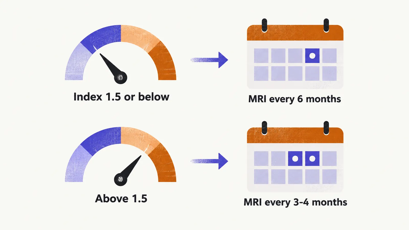
Risk stratification uses three factors: JC virus antibody status, treatment duration beyond about 2 years, and prior immunosuppressant exposure. When all three combine on natalizumab, risk exceeds 1 in 100. Antibody status alone is graded by the antibody index. An index above 1.5 marks markedly higher PML risk beyond 2 years of natalizumab: about 8.83 per 1,000 during months 25 to 48, against about 1.13 per 1,000 at index 1.5 or below.
The index guides MRI rhythm, not a stop rule. A baseline scan precedes natalizumab initiation. Patients at index 1.5 or below need MRI at least every 6 months. Patients above 1.5 need MRI every 3 to 4 months after 18 months of treatment, including diffusion-weighted sequences. The antibody index is retested serially because serostatus can change. Two cautions bound the test. Its sensitivity is high but its specificity is low: about 57% of patients who never develop PML also test above 1.5. A positive result therefore manages surveillance intensity and never by itself contraindicates treatment.
Vaccine timing around disease-modifying therapies
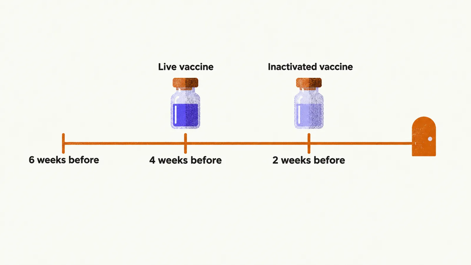
Immunization status is reviewed at diagnosis, before any immunosuppressive therapy starts, because immunosuppression blunts the response to vaccines and makes live vaccines risky. Inactivated vaccines can be given at any time, but ideally at least 2 weeks before treatment onset so the immune response completes. Live attenuated vaccines need at least 4 weeks before treatment onset, and 6 weeks before ocrelizumab or alemtuzumab.
Once immunosuppression is established, live vaccines are avoided on sphingosine-1-phosphate modulators and anti-CD20 therapies, and before immune reconstitution after cladribine or alemtuzumab. They are ideally avoided on natalizumab, dimethyl fumarate, and teriflunomide as well. After stopping an immunosuppressive therapy, live vaccines wait for a safety interval that ensures immune restoration. A short pulse of high-dose steroids postpones live vaccines by 1 month. Vaccination is also deferred during a relapse until the episode resolves or stabilizes.
Two special timings recur in practice. For anti-CD20 infusions given every 6 months, inactivated vaccines fit best at least 3 months after the last infusion and 4 to 6 weeks before the next one. Patients without measles or varicella immunity who face a plausible exposure seek immediate medical advice, because post-exposure immunoglobulin may be offered.
The standing vaccines are yearly influenza and pneumococcal vaccination for candidates for immunosuppressive therapy and for patients with significant disability. Household and close contacts should be immunized too, since the patient may respond partially or cannot receive live vaccines. Blunted responses are expected on some therapies: patients on sphingosine-1-phosphate modulators, anti-CD20 agents, alemtuzumab, or cladribine receive counseling that protection may be incomplete and that other precautions still apply.
Pregnancy planning
Pregnancy planning starts before conception, with the neurologist reviewing the disease-modifying therapy. Per-agent washout and contraception intervals differ, so each interval follows the current product label rather than any memorized table. Live vaccines that are still missing are completed at least 1 month before pregnancy, because live vaccines are contraindicated once pregnancy begins.
During pregnancy, inactivated vaccines are considered safe. Influenza vaccination is recommended in any trimester at the start of the influenza season. Pertussis protection (diphtheria, tetanus, pertussis) is advised in each pregnancy, preferably between weeks 20 and 36, to maximize antibody transfer to the newborn. Women without rubella or varicella immunity are identified during pregnancy and vaccinated postpartum before any disease-modifying therapy restarts.
Two newborn rules apply. Infants exposed to anti-CD20 therapy during or shortly before pregnancy have their CD19-positive B cells measured, and live vaccines such as rotavirus wait until B-cell recovery. Breastfeeding is compatible with vaccination except for yellow fever vaccine.
Laboratory monitoring principles
Each drug carries its own laboratory monitoring schedule, and the schedule follows the current product label. Dimethyl fumarate shows how such a schedule works, because lymphopenia (a low lymphocyte count) is its main laboratory signal. Lymphocyte counts are checked before starting and every 3 months during treatment. Sustained moderate lymphopenia, between 0.5 and 0.8 times 10⁹ per liter for longer than 6 months, prompts a reassessment of benefit against risk. Counts below 0.5 times 10⁹ per liter persisting beyond 6 months prompt discontinuation. MRI vigilance increases for patients at higher PML risk, in line with local recommendations.