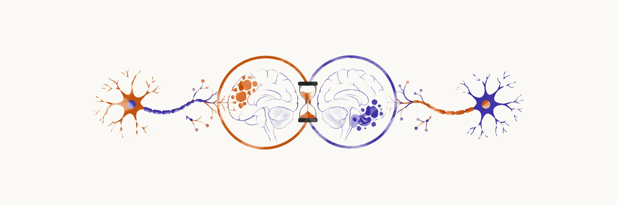
MS is a clinical diagnosis supported by investigations. Every version of the McDonald criteria asks the same two questions in a patient with a compatible presentation: are lesions scattered in separate places (dissemination in space), and is the disease ongoing over time (dissemination in time), while alternative diagnoses are excluded. The criteria exist to prevent both over-diagnosis, which commits someone to costly and potentially harmful treatment, and under-diagnosis, which lets silent disease progress untreated.
Dissemination in space and time
Dissemination in space is shown by lesions in more than one characteristic area of the nervous system, and it is counted on MRI as T2 lesions (bright foci on T2-weighted images) in defined locations. Under the 2017 criteria, it requires at least 1 T2 lesion in at least 2 of 4 areas: periventricular (next to the lateral ventricles), cortical or juxtacortical (in or beside the cortex), infratentorial (brainstem and cerebellum), and spinal cord. Under the 2024 criteria, five locations count: periventricular or subcortical, infratentorial, cortex, spinal cord, and optic nerve, with two required.
Dissemination in time can be shown by the simultaneous presence of enhancing and non-enhancing asymptomatic lesions on one scan, meaning some plaques are new and others old, or by a new T2 or enhancing lesion on any follow-up MRI. Since 2017, oligoclonal bands may substitute for this requirement.
How the criteria evolved
The 2010 criteria demonstrated dissemination in space and time mainly through clinical attacks and MRI. For progressive onset from the start, they required 1 year of disability progression plus two of three: positive brain MRI, positive spinal cord MRI, or positive cerebrospinal fluid.
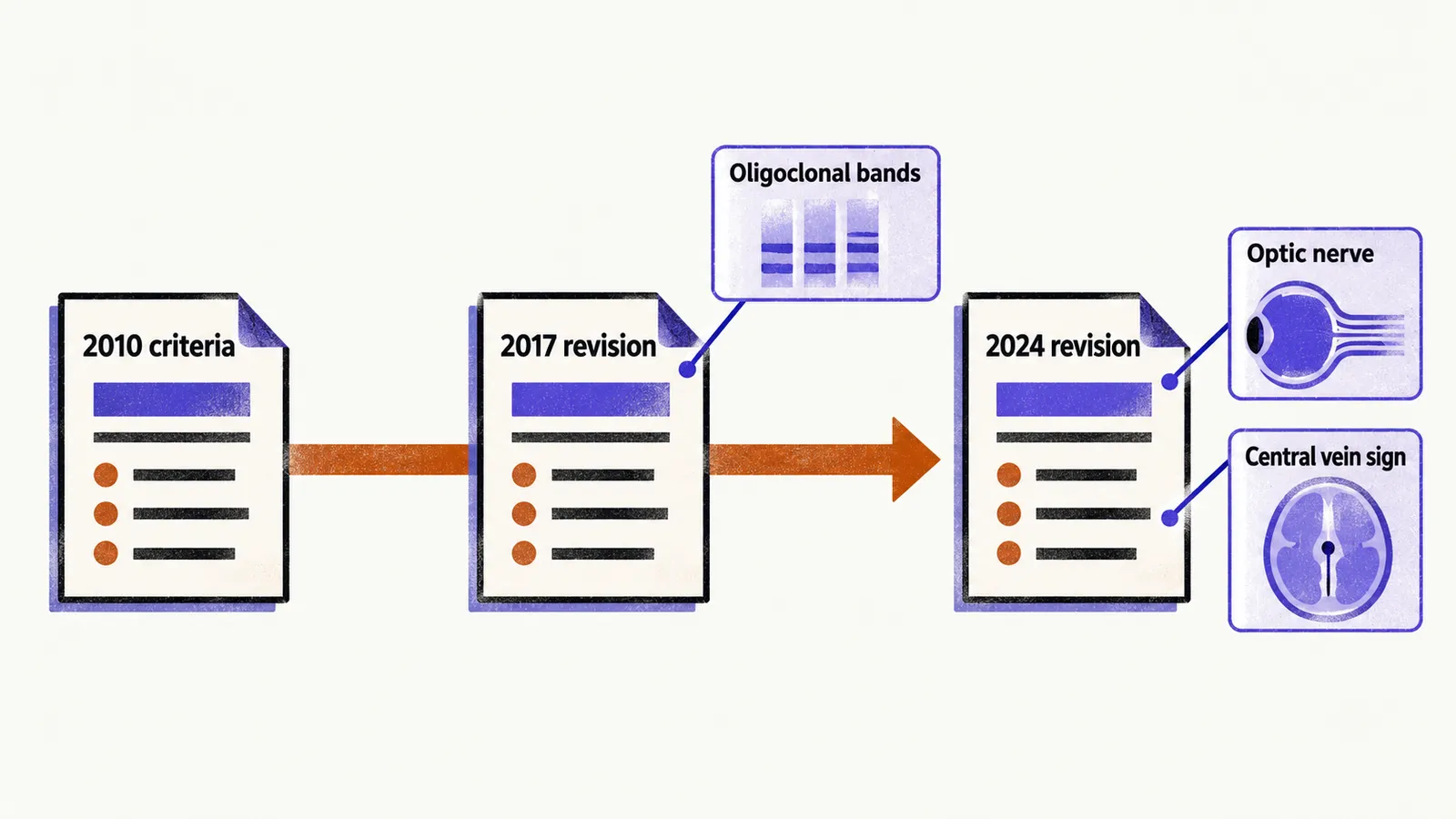
The 2017 revision made two changes. It added cortical and juxtacortical lesions to the dissemination-in-space locations, and it allowed cerebrospinal-fluid-specific oligoclonal bands to substitute for dissemination in time. A patient with a single attack and a single MRI could therefore be diagnosed without waiting for a second scan, which moved treatment earlier.
The 2024 revision shifted emphasis further. The optic nerve joined the dissemination-in-space locations, bringing the count to five topographies, of which two must show typical lesions. Lesion characteristics gained diagnostic weight: the central vein sign and paramagnetic rim lesions can now support the diagnosis in patients with typical presentations who do not fully meet classical dissemination criteria. The stated aim is a diagnosis that is easier and faster, so that treatment starts before further irreversible injury.
Cerebrospinal fluid
Two markers indicate antibody production inside the nervous system, which matters because it can substitute for dissemination in time. Oligoclonal bands detected by isoelectric focusing show a pattern present in cerebrospinal fluid but not serum, and reported figures give roughly 93% sensitivity with 94% specificity. The Link index, the ratio of cerebrospinal-fluid-to-serum IgG divided by the cerebrospinal-fluid-to-serum albumin ratio, rises above about 0.7 when intrathecal IgG synthesis is present. Earlier criteria accepted either marker; the 2017 criteria focus on cerebrospinal-fluid-specific oligoclonal bands.
Evoked potentials and emerging markers
Imaging and cerebrospinal fluid are not the only ways to show that the disease is present. Multimodal evoked potentials (visual, auditory, somatosensory), which record the brain’s electrical response to a sensory stimulus, can reveal clinically silent conduction slowing. The characteristic pattern is increased latency (delay) with preserved amplitude (signal size), meaning conduction is slowed by demyelination without complete axonal interruption. Abnormal visual evoked potentials also predict conversion from radiologically isolated syndrome to clinical disease.
Neurofilaments, structural proteins of axons released when axons are damaged, are an emerging marker of ongoing neurodegeneration in serum and cerebrospinal fluid. Sensitive assays may eventually help predict progression and monitor treatment response, but threshold values for routine diagnostic use are not established.
Red flags
Because MS is a diagnosis of exclusion as much as of inclusion, the clinician also looks for features that argue against it. Clinical red flags include absent ocular involvement, absent corpus callosum or spinal cord lesions, very early or very late onset, no recovery between episodes, disease manifestations outside the nervous system, and a family history pointing to a different neurological disorder. Atypical MRI patterns similarly demand reconsideration. Before committing to disease-modifying therapy, particularly with an optico-spinal presentation, testing for aquaporin-4 and myelin oligodendrocyte glycoprotein antibodies is standard practice to exclude neuromyelitis optica spectrum disorder and myelin oligodendrocyte glycoprotein-associated disease.