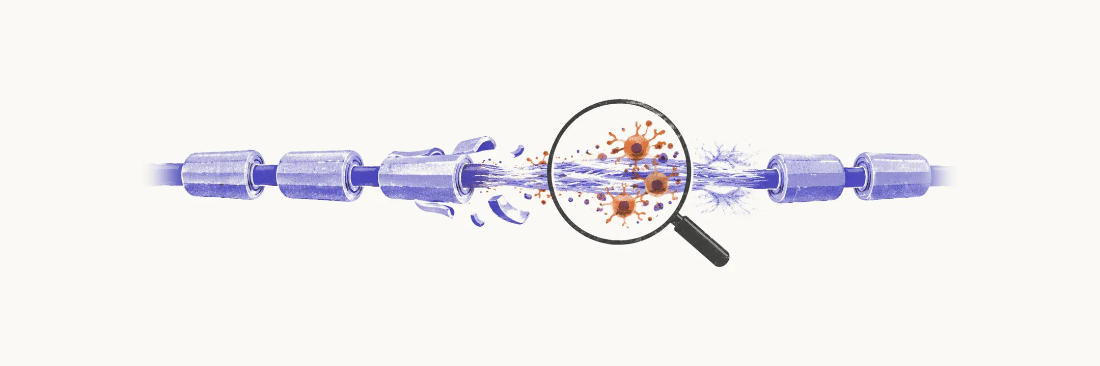
An MS plaque is a focal area where myelin has been destroyed by inflammation, with variable injury to the axons running through it. Three processes define it: demyelination, inflammation, and axonal loss. Early lesions show myelin destruction with relative axonal preservation, while older lesions add irreversible axonal loss and scarring. This progression explains the two kinds of disability in MS. Relapse-related disability tracks with inflammation and demyelination, which can partly recover, while progressive disability tracks with axonal loss, which cannot.
Active versus chronic plaques
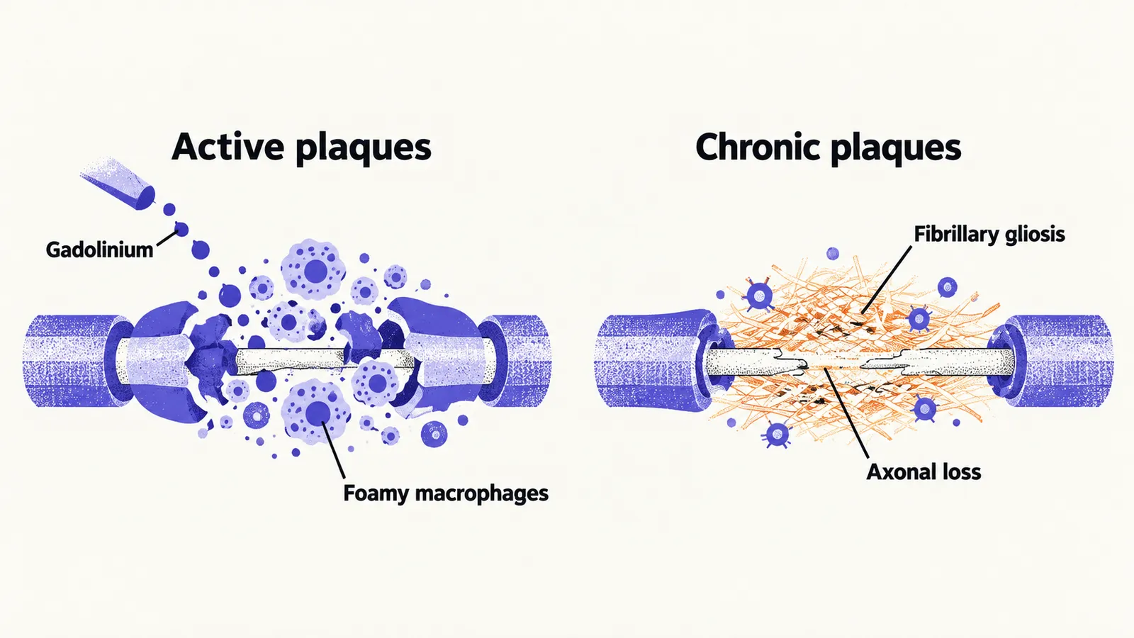
Active plaques are hypercellular, meaning crowded with cells. They contain foamy macrophages digesting myelin, reactive astrocytes (star-shaped glial cells that respond to injury), and perivenular inflammation around small veins, with active myelin breakdown advancing outward at the lesion edge. Because the blood-brain barrier is disrupted where inflammation is active, these lesions enhance with gadolinium, a contrast agent that leaks into tissue, on MRI.
Chronic plaques are sharply demarcated and hypocellular, meaning sparsely populated by cells. The dominant feature is fibrillary gliosis, a glial scar, with axonal damage and loss and no active myelin breakdown. Mature oligodendrocytes, the cells that make and maintain myelin, are markedly reduced or absent, which is why chronic lesions remyelinate poorly even after inflammation subsides. Some perivascular inflammation may persist, and partial remyelination can sometimes be found at the edge.
Where plaques sit
Typical locations are the periventricular white matter, optic nerves and chiasm, corpus callosum, brainstem, cerebellum, and spinal cord, especially the cervical segment. The perivascular distribution follows from the outside-in pathogenesis: inflammatory cells arrive through small veins and spread outward along perivenular spaces. Gross examination in advanced disease may show brain atrophy with widened cortical sulci, enlarged ventricles, and mild spinal cord swelling with thickened meninges, all reflecting cumulative tissue loss.
Injury beyond the plaques
Axonal injury begins early. Damaged axonal terminals and exposed neurofilaments are already visible during predominantly inflammatory phases, and neurodegeneration appears to precede the first clinical episode. Areas of so-called dirty-appearing white matter show diffuse myelin and axonal loss without forming discrete plaques, representing disease burden beyond the counted lesions. Which symptoms a patient develops depends on where in the nervous system the plaques sit.