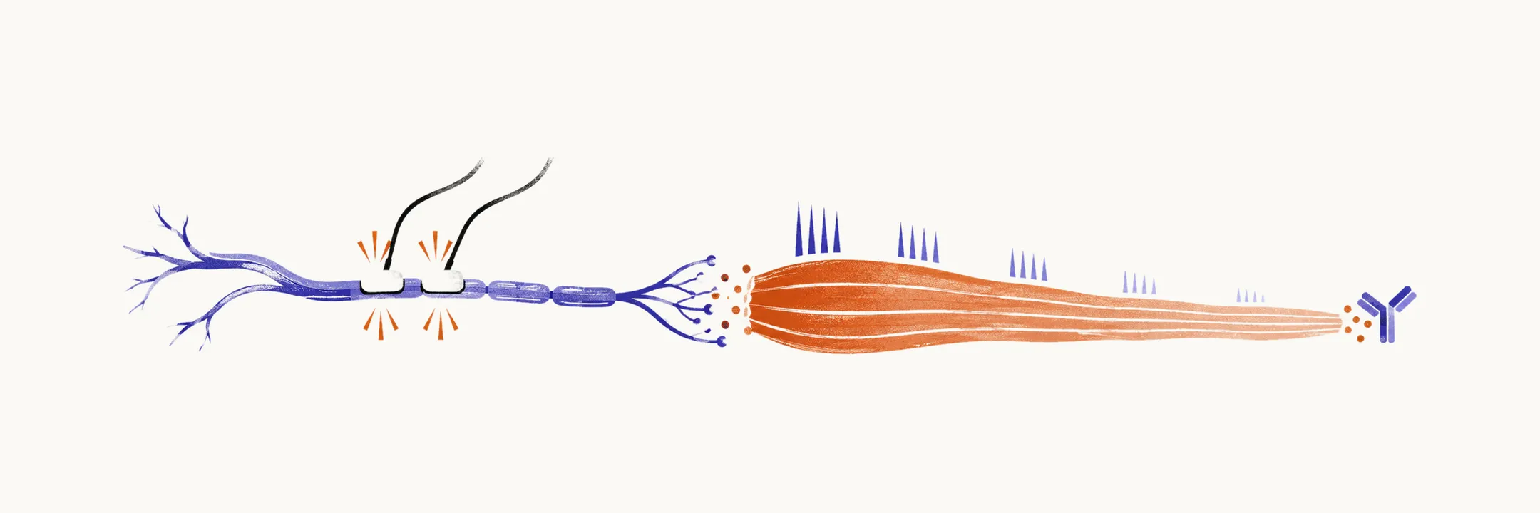
A patient with myasthenia gravis, an autoimmune disorder of transmission at the neuromuscular junction where nerve meets muscle, can look entirely normal during a brief examination because the weakness fluctuates and recovers with rest. Diagnosis therefore means provoking the weakness first, then proving it sits at the neuromuscular junction rather than in muscle, nerve, or brain.
Provoking weakness at the bedside
Fatiguing manoeuvres outlast the patient’s reserve and expose what rest conceals:
- Sustained upgaze for about 30 seconds — watch for emerging or worsening ptosis and diplopia.
- Sustained lateral gaze for about 30 seconds — produces or worsens diplopia from extraocular fatigue.
- Arm abduction against gravity — watch for progressive drooping of the held limbs.
- Counting aloud on one breath — the voice fades, turns soft and indistinct, then recovers after rest, testing bulbar and respiratory reserve.
- Repetitive movements such as clapping or sit-to-stand — count how many repetitions precede visible weakening.
Ocular motility documentation, for example with the Lancaster test of gaze positions, records the extraocular pattern in ocular disease.
The time of examination matters. A morning visit may miss disease that an evening visit reveals, and spontaneously resolving ptosis (a drooping eyelid) does not mean the disease has gone.
Bedside pharmacological clues
Beyond fatiguing manoeuvres, bedside tests can exploit how the junction handles acetylcholine. The ice pack test is the most useful bedside confirmatory manoeuvre for ptosis: cooling the drooping eyelid for about 2 minutes produces transient improvement. Cold inhibits acetylcholinesterase, the enzyme that breaks down acetylcholine at the junction, so acetylcholine lingers longer and stimulates the remaining receptors. The improvement fades soon after the ice is removed, and the test is non-invasive and safe.
Edrophonium, a short-acting acetylcholinesterase inhibitor given intravenously, briefly but dramatically restores strength; because of cardiac risks it is used only with monitoring and has largely receded from routine practice. An oral pyridostigmine trial over days, typically starting from 60 mg doses, can support the diagnosis when symptoms improve with regular dosing, doubling as initial therapy rather than a rapid test.
Electrodiagnosis
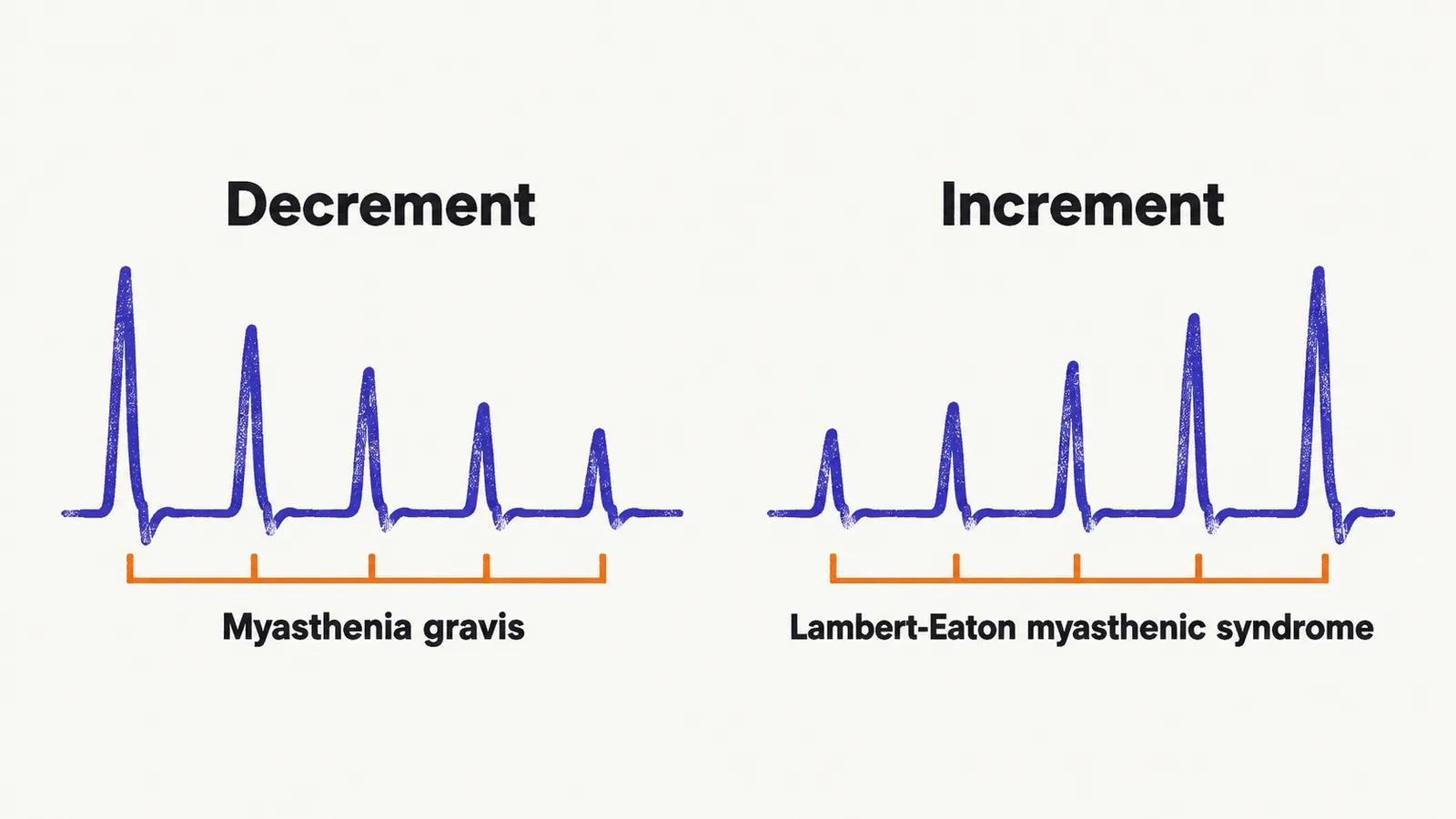
Repetitive nerve stimulation is the standard electrical test. A motor nerve is stimulated at low frequency, usually 2–3 Hz, and the muscle response amplitude is recorded across successive stimuli. In myasthenia gravis the amplitude decrements, typically by more than 10% between the first and fourth or fifth response, because each impulse finds fewer functional receptors. The mirror image, an incremental response to rapid stimulation or after brief exercise, points to Lambert-Eaton myasthenic syndrome, a presynaptic disorder that mimics myasthenia gravis, as detailed in MG versus LEMS.
Single-fibre electromyography is the most sensitive test of junction transmission. A fine electrode records two fibres of one motor unit: the interval between their activations (jitter) widens, and some impulses fail entirely (blocking). Sensitivity comes at the cost of specificity, since any transmission defect can disturb the recording, so the result is read together with the clinical picture.
Antibodies
Antibody testing confirms the disease and predicts its behaviour. Anti-acetylcholine-receptor antibodies are tested first: highly specific at about 99.8%, meaning almost no false positives, and positive in roughly 85% of generalised and half of ocular cases (Gilhus et al., 2019). A positive result in the right clinical context confirms the diagnosis without further workup.
When receptor antibodies are negative, anti-MuSK testing follows. MuSK-positive disease is predominantly bulbar with minimal ocular signs, responds poorly to pyridostigmine and thymectomy, and responds well to rituximab, so the result changes management. Anti-LRP4 antibodies explain a fraction of double-negative cases and usually behave mildly. The remaining 5–10% are seronegative for all three and are diagnosed clinically with electrodiagnostic support; research antibodies against agrin, cortactin, and related junction proteins may eventually explain some of these cases. Results take days to weeks, so bedside manoeuvres guide immediate decisions while the results are pending.
Imaging
Chest computed tomography (CT) is mandatory in every newly diagnosed patient to screen the thymus, the gland whose tumour or overgrowth is linked to the disease: thymoma in 10–15% and hyperplasia in roughly 70% of non-thymomatous cases. Thymoma can extend into pericardium, pleura, or beyond, and its presence mandates removal.
Workup sequence in practice
In practice the steps run in this order:
- Bedside fatiguing manoeuvres once fatigable, fluctuating weakness is suspected.
- The ice pack test when ptosis is present.
- The antibody panel, starting with anti-receptor antibodies.
- Electrodiagnosis to confirm the junction defect.
- Chest imaging for every confirmed case.
A confirmed diagnosis, its antibody subtype and the thymic findings together shape which treatment is chosen.