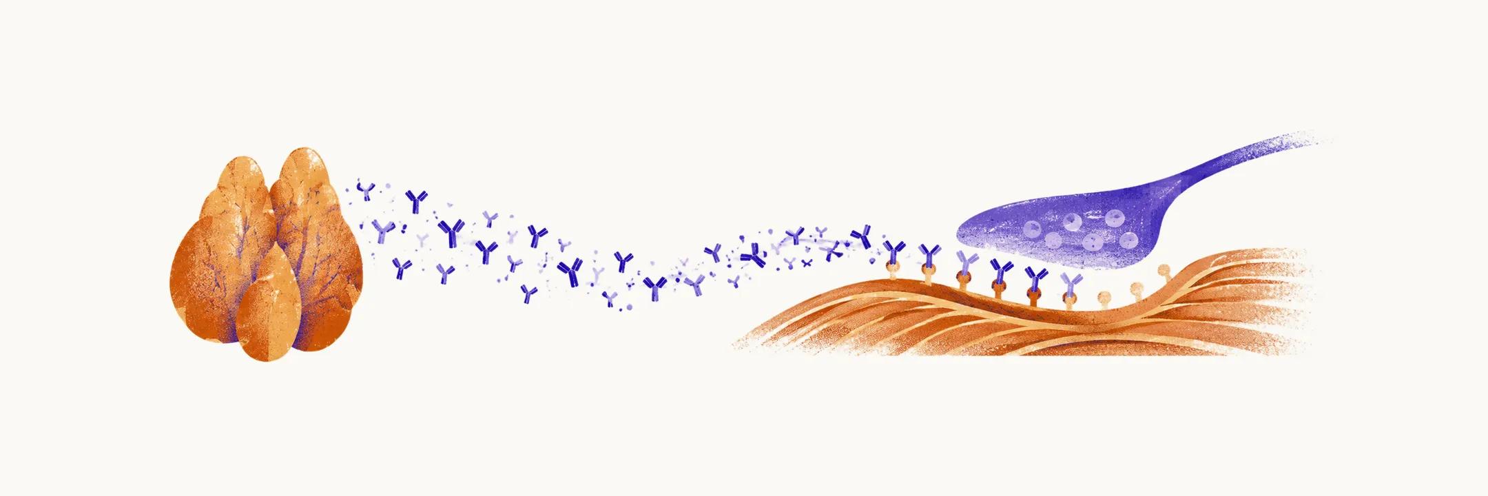
Myasthenia gravis is an autoimmune disease in which antibodies attack the endplate, the postsynaptic membrane of the neuromuscular junction that belongs to the muscle.
The junction in health
A motor nerve ending releases acetylcholine into the synaptic gap, and receptors on the folded muscle membrane catch it and trigger contraction. The receptors are densely clustered on the folds, which gives transmission a wide safety margin: more acetylcholine is released than the receptors need to depolarise the fibre. Acetylcholinesterase in the gap breaks the transmitter down and so ends the signal.
What the antibodies do
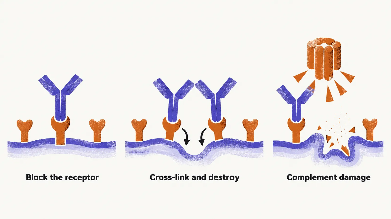
In myasthenia gravis the release is normal but fewer receptors answer. Antibodies against the acetylcholine receptor damage the endplate in three ways: they block the receptor’s acetylcholine-binding site, they cross-link receptors so that the muscle cell takes them in and destroys them, and they activate complement, the immune cascade that damages the folded membrane holding the receptors in place. Receptor antibodies belong mainly to the IgG1 and IgG3 subclasses of immunoglobulin G, the main circulating antibody class; these subclasses activate complement.
With fewer receptors, the safety margin narrows. A single impulse may still trigger a contraction, but each repeated signal finds less reserve, so weakness grows with use and eases with rest. That is what makes the weakness fatigable.
Antibody subtypes
Most generalised disease is driven by antibodies against the acetylcholine receptor itself, found in roughly 85% of generalised cases and about half of purely ocular cases (Gilhus et al., 2019). Patients who lack these are called seronegative for receptor antibodies, and the other subtypes cluster among them.
Antibodies against muscle-specific tyrosine kinase (MuSK), a protein that clusters receptors at the membrane, produce a distinctive pattern: predominantly bulbar weakness (of the speech and swallowing muscles) with minimal ocular involvement, sometimes isolated head drop, a poor response to pyridostigmine (a drug that raises acetylcholine levels in the gap) and thymectomy (surgical removal of the thymus), and a good response to rituximab (a drug that depletes antibody-producing B cells). MuSK antibodies are mainly IgG4, which does not activate complement, and they disrupt the receptor-clustering pathway instead of damaging the membrane directly (Lazaridis & Tzartos, 2020).
LRP4 is the muscle receptor for agrin, the nerve-derived signal that activates MuSK; antibodies against it account for a fraction of double-negative cases (negative for both receptor and MuSK antibodies) and usually behave mildly. Across all patients, MuSK antibodies are found in roughly 6% and LRP4 antibodies in about 2% (Lazaridis & Tzartos, 2020). About 5–10% of patients are seronegative for all three and are diagnosed on clinical grounds plus electrodiagnosis.
Antibody levels do not track severity in a strict dose–response way, so a modest titre never excludes severe disease. A falling titre during treatment generally parallels clinical improvement.
The thymus connection
The thymus, a gland in the chest where T cells mature, is where the autoimmune response is organised: thymic tissue expresses receptor-like proteins that activate T cells, which in turn drive B cells to produce the circulating antibodies. About 10–15% of patients have a thymoma, a thymic tumour, which can invade locally and must be removed regardless of its apparent contribution to the weakness.
Around 70% of patients without thymoma show lymphoid follicular hyperplasia of the thymus instead, an overgrowth of lymphoid follicles. Because the thymus sustains the antibody response, thymectomy lowers subsequent immunosuppressive requirements, with benefit most firmly established in acetylcholine-receptor-positive generalised disease without thymoma (Wolfe et al., 2016). It is generally not recommended in MuSK-positive disease, where the pathology is not thymus-driven.