Diarrhea is not one disease. It is the shared end point of several different failures in intestinal water handling, and the mechanism matters because it predicts the stool electrolyte pattern, the response to dietary change, and the way the child should be rehydrated.
How the intestine normally handles water
Everything swallowed meets a continuous stream of endogenous fluid: saliva, gastric juice, bile, pancreatic juice and intestinal secretions. Together these make up a large daily volume that enters the small intestine, and under normal circumstances almost all of it is reabsorbed before the stool. Only a small amount of water normally leaves the body this way.
Diarrhea therefore means reabsorption has failed. There are three ways this can happen: more solute has been left in the lumen (the inside of the bowel) than the mucosa (its lining) can carry; secretion into the lumen has overtaken absorption; or the absorbing epithelium, the surface layer of cells, has itself been damaged. The last of these is the commonest route in children with acute gastroenteritis.
Osmotic and secretory diarrhea
Osmotic diarrhea occurs when an osmotically active solute — a dissolved substance that holds water on its side of the lining — remains in the intestinal lumen and keeps water with it. Lactulose, a sugar that normal mucosa does not absorb, is the clean prototype. In viral gastroenteritis the same logic applies through damage. The virus injures the small-bowel epithelium, and the brush-border enzymes — digestive enzymes on the surface of those cells — including lactase, fall. Unabsorbed carbohydrate stays in the lumen, and water follows it. The mucosa is anatomically abnormal, so digestive and absorptive function is compromised as well.
Secretory diarrhea occurs on an intact mucosa. An enterotoxin (a toxin that acts on intestinal cells) or an endogenous secretagogue (a substance made by the body that stimulates secretion) raises intracellular second messengers, the signalling molecules inside the cell. The epithelium then does two things at once: it secretes chloride and water into the lumen and it stops absorbing sodium. Cholera is the classical example. The mucosa looks normal under the microscope — the transport behaviour of the cell has changed, not its structure.
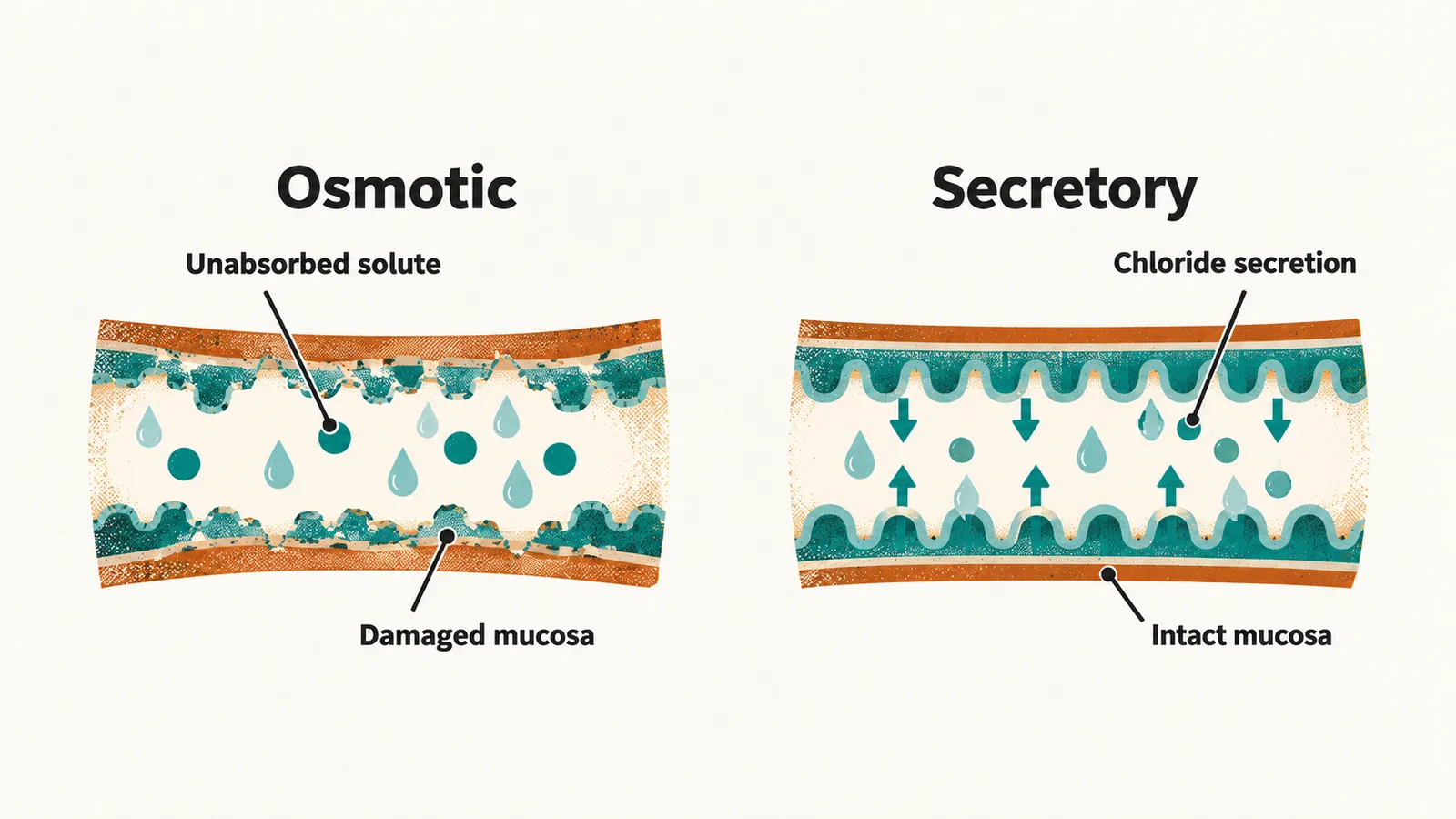
The clinical consequence of the split is that the two heal differently. An osmotic diarrhea settles when the offending solute is removed or the epithelium regenerates, which is why dietary decisions matter in viral disease. A secretory diarrhea settles when the toxin is cleared, with no structural repair to wait for.
What stool electrolytes show
Stool fluid normally has an osmolality — a concentration of dissolved particles — close to that of plasma, because whatever is left in the lumen equilibrates with plasma water. That stays true when unabsorbed solute is holding water in the lumen; what changes is the makeup of the osmoles, with sodium and potassium accounting for only a small share of the total. When secretion is driving the loss, by contrast, sodium and bicarbonate appear in the stool in far higher concentrations than usual.
The fecal osmotic gap formalises this. It is estimated as plasma osmolality minus twice the sum of stool sodium and potassium. A large gap means that much of the stool’s osmolality comes from something other than sodium and potassium, which points to an osmotic mechanism; a small gap points to a secretory one. The gap can be calculated at the bedside in watery diarrhea, but it belongs to the chronic or unexplained picture more than to ordinary acute gastroenteritis, where the mechanism is usually apparent from context.
| Feature | Osmotic | Secretory |
|---|---|---|
| Mucosa | Damaged | Intact |
| Usual cause in children | Viral enteritis with brush-border enzyme loss | Enterotoxin, as in cholera |
| Stool electrolytes | Sodium low relative to plasma | Sodium high, close to plasma |
| Fecal osmotic gap | Large | Small |
| What resolves it | Removing the solute, or mucosal healing | Clearance of the toxin |
Where dehydration comes from
Whatever the mechanism, the water that is not reabsorbed is lost from the body, and that loss is what makes diarrhea dangerous. Dehydration is a loss of body water with a matching fall in plasma volume. Children reach it sooner than adults: they turn over more water per kilogram, have a larger surface area relative to weight, lose more through the skin and lungs, and in early infancy concentrate urine less effectively. Acute diarrhea is the most common cause of dehydration in early childhood. Four routes produce it:
- Gastrointestinal loss — the dominant route in children, of which acute gastroenteritis is the prototype.
- Renal loss — osmotic diuresis in diabetes, free-water loss in diabetes insipidus, adrenal insufficiency, chronic renal impairment.
- Cutaneous loss — burns.
- Insensible loss — fever and tachypnea increase what is lost through skin and lungs without anything visible being lost.
Once volume has fallen, two further things follow. Sodium concentration in plasma decides whether the deficit is being replaced with a relatively sodium-poor or sodium-rich fluid, and therefore how quickly sodium may safely be corrected; recognising the sodium pattern belongs to Clinical Assessment of Acute Diarrhea and Dehydration, and correcting it to Rehydration Management in Acute Diarrhea. The mechanism, in turn, decides when the gut is ready to be fed again, and the dietary consequences of a damaged brush border belong to Refeeding and Drug Therapy in Acute Diarrhea.
In children, the usual causes line up with the two mechanisms: viral enteritis that injures the small-bowel epithelium produces the osmotic picture, and an enterotoxin acting on an intact mucosa produces the secretory one. Knowing which organisms are common, and the pattern each produces, is therefore the next step in reading a child with acute diarrhea.