Acromegaly is suspected from its clinical features, confirmed biochemically with insulin-like growth factor 1 (IGF-1) and the oral glucose tolerance test, and assessed by imaging of the pituitary. Treatment is mainly surgical resection of the adenoma, with radiotherapy and drug therapy as further options.
When to suspect acromegaly
The panel for the diagnosis of acromegaly is initiated in two main clinical scenarios. The first is evident and overt clinical manifestations related to acromegaly, such as acral facial features, enlargement of the hands and feet and dental diathesis (interdental separation), which are common in overt acromegaly cases. The second is intermediate changes that could be associated with acromegaly, in which the disease is still in the early stages and clinical diagnosis would prevent many complications in the future; these patients mostly manifest hypertension, carpal tunnel syndrome, sleep apnea, hyperhidrosis, debilitating arthralgia, refractory headache or amenorrhea.
Because the change is slow, the patient can be asked to bring photographs from different years: by the chronic and long-term effect of GH on the body, an obvious change in the shape of the face may be seen.
Biochemical diagnosis
Diagnosis of acromegaly is made by combining clinical examination with biochemical tests. Due to the pulsatility of GH, GH cannot be used by itself to diagnose GH hypersecretion disorders, and the level of GH is not correlated with the severity of disease. The guideline recommends screening with IGF-1 and confirmation of the diagnosis with the oral glucose tolerance test.
IGF-1 level
A single IGF-1 (insulin-like growth factor 1) level is considered the first line of biochemical test for the diagnosis of acromegaly. The level of IGF-1 in the plasma depends on several physiological and pathological factors: physiologically on age and gender, and pathologically on liver dysfunction, renal dysfunction, nutrition and DM (diabetes mellitus).
GH day curve
For assessment of GH hypersecretion, serial sampling can be performed: 5 samples over 12 hours. The 5 samples for measurement of the mean GH level should be taken from an indwelling venous cannula, to prevent the effect of the stress of puncturing during sampling, because GH secretion is extremely sensitive to physiological stressors such as exercise; random sampling can be useless due to the overlap that can occur between normal subjects with physiological stress and acromegalic patients.
The IGF-1 level is not affected by physiological stress and is therefore a better option for evaluation of acromegalic patients; in fact, the GH level fluctuates in these patients, but the IGF-1 level is constantly high.
Random sampling of GH is useful for exclusion of the diagnosis of acromegaly in only one condition: GH < 0.4 ng/ml together with a normal IGF-1, based on age and sex.
Oral glucose tolerance test
In physiological conditions the serum level of GH is suppressed after the ingestion of glucose, and failure of this physiological response can occur in GH hypersecretion. The gold-standard diagnostic test for GH hypersecretion diseases is the oral glucose tolerance test (OGTT).
To perform the OGTT: oral ingestion of 75 gram of glucose at 9 a.m. in a fasted patient, with serial sampling at baseline and then 30 minutes, 60 minutes, 90 minutes and 120 minutes after the ingestion.
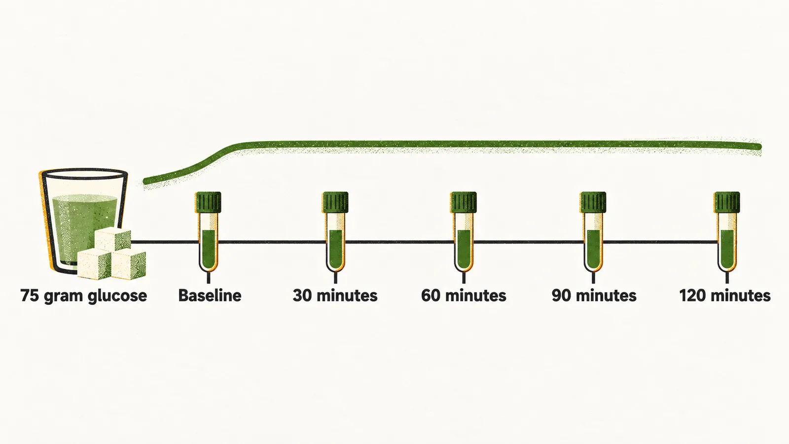
In normal subjects the level of GH after the OGTT becomes undetectable, or less than 0.1 ng/ml, while in acromegaly patients it remains detectable, and in about 30% of the cases there is a paradoxical increase of GH. Failure to suppress the GH level to < 0.4 ng/ml supports the diagnosis of acromegaly. With older assays (the laboratory methods used to measure GH) the guideline threshold for normal suppression was a GH nadir (lowest value) below 1 ng/ml, and the lower 0.4 ng/ml cutoff applies to modern, more sensitive assays.
False-positive results of the OGTT can be seen in:
- stressful conditions
- DM
- cirrhosis
- adolescence
- chronic renal failure
- use of L-dopa and estrogen
- heroin addiction
TRH test
Thyrotrophin-releasing hormone (TRH) injection physiologically inhibits the release of GH. 200 μg of intravenous TRH, with serial measurement at baseline and 20 and 60 minutes after the injection, is suggested in patients with mild acromegaly with a low level of circulating GH, to confirm the diagnosis.
Other tests
IGFBP3 (insulin-like growth factor binding protein 3), GHRH (growth hormone-releasing hormone) and prolactin may also be measured.
Pituitary imaging and further workup
The optimal modality for investigation of the pituitary gland in patients with suspected acromegaly is MRI with gadolinium enhancement, for defining the tumor size and characteristics and possible invasion of surrounding structures, such as cavernous sinus invasion and suprasellar extension.
Neuro-ophthalmological assessment should be done in all patients with radiological evidence of a macroadenoma, especially in the case of optic contact by the tumor. Its purpose is assessment of visual complications such as optic atrophy, retinal vein engorgement and papilloedema due to compression.
Histological studies that can be performed on the somatotrope tumors are:
- distinguishing between sparsely and densely granulated tumors, by use of keratin staining
- Ki-67 index
- immunohistochemistry of Pit-1, SF-1 and Tpit
- tumor response to somatostatin analogue therapy, by means of somatostatin receptors (SSTR)
Secondary hypopituitarism
Pituitary function is the other follow-up that matters once the diagnosis is made. Secondary hypopituitarism mostly occurs in the case of macroadenomatous acromegaly; it occurs in 15–20% of the cases, and can be secondary central hypothyroidism, secondary central hypogonadism or secondary central hypoadrenalism.
Differential diagnosis
Tall stature raises the suspicion of acromegaly in most cases, but when soft tissue and biochemical evidence are absent, the differential diagnosis is:
- Marfan syndrome
- homocystinuria
- familial trait
Screening for complications
Acromegaly is associated with quite a lot of important comorbidities that are present at the time of diagnosis; therefore assessment for possible complications is suggested in patients with acromegaly at the time of confirmation of the diagnosis. In the table, BP is blood pressure, OSA obstructive sleep apnea, HbA1c glycated hemoglobin, FBG fasting blood glucose, US ultrasound, DEXA dual-energy X-ray absorptiometry, and QOL quality of life, which ACROQOL assesses.
| Suggested Strategy for Screening for Complications | ||
|---|---|---|
| Co-morbidity to evaluate | Screening test | Frequency |
| Hypertension | Measurement of BP; ambulatory BP monitoring in selected cases | 6 monthly |
| DM | HbA1c, FBG; OGTT in selected cases | At diagnosis and 6 monthly if normal |
| OSA | Clinical evaluation and Epworth scale; polysomnography for confirmation | At diagnosis and annually |
| Cardiomyopathy | ECG; echocardiography | At diagnosis and 3–5 yearly if normal |
| Dyslipidemia | Lipid profile | At diagnosis and 6 monthly |
| Colon polyps | Colonoscopy | At diagnosis in patients >40 years of age, and 10 yearly if normal, 3–5 yearly if polyp noted and IGF-1 elevated |
| Thyroid nodules | Clinical evaluation; thyroid US guided by examination | Annually |
| Vertebral disease | Bone morphometric study using thoracic x-ray, thoracic and lumbar spine x-ray; DEXA | At diagnosis and yearly, guided by symptoms |
| Cerebral aneurysm | Cerebral MR angiography | Infrequently |
| QOL | ACROQOL | Annually |
Treatment
Treatment of patients diagnosed with acromegaly is mainly based on surgical resection of the adenoma as the first line of treatment; the second line of treatment is radiotherapy. The aim is to normalize IGF-1 and suppress GH, because it is uncontrolled disease that drives the excess mortality of acromegaly.
Surgical resection is usually performed by the transsphenoidal route. The chance of remission depends on the size and invasiveness of the tumor: average cure rates are about 78% for microadenomas (less than 1 cm) and 50% or less for macroadenomas (1 cm or more).
Radiotherapy, the second line of treatment, has a slow effect, often taking years to bring GH and IGF-1 under control, and it carries a risk of hypopituitarism.
The pharmacological options that can be used in these patients are dopamine agonists, somatostatin analogues and GH-receptor antagonists. Somatostatin analogues (such as octreotide and lanreotide), which act mainly on somatostatin receptor type 2, are the first-line medical option when surgery is not curative or not possible; as monotherapy they normalize IGF-1 in only about 40% of patients. Pegvisomant is a GH-receptor antagonist used as a second-line agent or combined with a somatostatin analogue, and the combination can normalize IGF-1 in most patients. Dopamine agonists such as cabergoline can be used, particularly when IGF-1 is only modestly elevated.