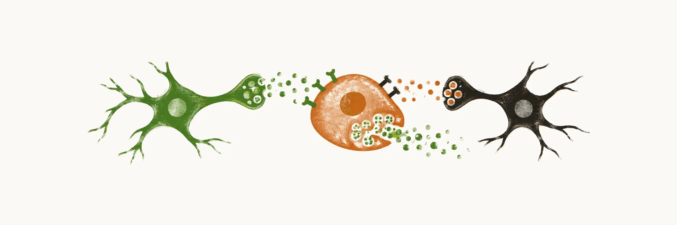
Growth hormone (GH) is a 191 amino-acid hormone with two disulfide bonds, made by the somatotrope cells of the pituitary gland, and it has a very high homology with the prolactin hormone.
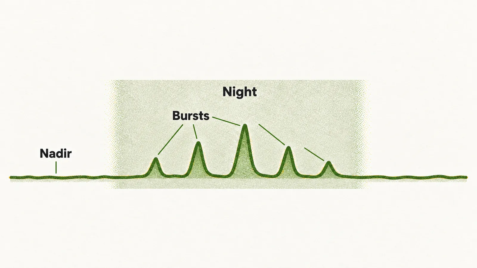
Growth hormone is not released steadily. Secretion comes in pulsatile bursts, 4–11 times a day, that mostly occur during the night, and between the episodes of burst secretion the plasma concentration of GH reaches its nadir (its lowest level). Because of this pattern of bursts and nadirs, a single GH measurement is difficult to interpret, and the GH concentration is not usually used on its own as a diagnostic tool.
Mediators of growth hormone release
The somatotrope cells are controlled by a balance of stimulatory and inhibitory signals, and some of these signals reach them through neurons of the hypothalamus.
The main stimulus is growth hormone-releasing hormone (GHRH); the neurons that secrete it are mostly located in the arcuate nucleus and the ventromedial nucleus of the hypothalamus. Ghrelin is a peptide that has a potent effect on GH secretion, expressed mainly by the gastrointestinal tract and the central nervous system, largely in response to hunger and hypoglycemia. Klotho is a further stimulatory mediator.
The inhibitory signal is the hypothalamic peptide somatostatin.
Dopamine, serotonin, neuropeptide Y and norepinephrine change GH release by acting on the GHRH-secreting and somatostatin-secreting neurons rather than on the pituitary directly. The rise of GH during fasting is attributed to neuropeptide Y.
Factors that change growth hormone secretion
The body’s energy state and its hormonal state both have an important effect on GH secretion.
On the hormonal side, children with hypothyroidism have lower levels of GH and IGF-1, which contributes to short stature. High glucocorticoids suppress GH secretion, so children with Cushing disease grow poorly.
Blood glucose acts in both directions. Hypoglycemia induced by insulin potently provokes GH secretion, and the increase is caused by inhibition of somatostatin release from the hypothalamus. Acute hyperglycemia, in contrast, produces a delayed GH increase: a healthy person can respond to hyperglycemia in two ways, inhibition of GH secretion from the pituitary gland and an increase in GHRH release from the hypothalamus, and the latter is the likely cause of the delayed rise.
Nutrients matter as well: high amino acids induce GH secretion, and high non-esterified free fatty acids (FFA) suppress it.
The table collects these and further physiological and pathological factors.
| PHYSIOLOGICAL | PATHOLOGICAL | ||
|---|---|---|---|
| Factors which increase GH secretion | Factors which reduce GH levels | Factors which increase GH secretion | Factors which reduce GH levels |
| Sleep | Overeating | Acute glucocorticoid excess | Chronic excess cortisol/glucocorticoids |
| Fasting | Obesity | Type 1 DM | Hyperthyroidism |
| Stress | Aging | Renal failure | Type 2 DM |
| Exercise | Increased IGF-1 | Acute use of opioids | |
| Hypoglycemia | Anorexia | ||
| Dopamine | Depression | ||
| Increased amino acids, i.e., a high-protein meal | Cirrhosis | ||
| Reduced free fatty acids | |||
| Glucagon | |||
| Testosterone and estradiol |
The growth hormone receptor and its signalling
GH acts through the growth hormone receptor (GHR), a receptor that is widely expressed in the body. In the plasma, GH is carried by a specific protein called GHBP (growth hormone-binding protein), which comprises the extracellular portion of the GHR.
The GHR is a tyrosine kinase receptor and signals only after dimerization: for activation, GH has to attach at the same time to two GHR molecules to form a dimerized molecule. Once the GH tyrosine kinase receptor has dimerized, several intracellular messengers are activated:
- STAT5, STAT3 and STAT1
- SRC, which activates MAPK
- IRS, which leads to PI3K and Akt
IGF-1
One of the major cell products made because of the effect of GH on the target cells is insulin-like growth factor 1, or IGF-1. It was classically believed to be made by the liver, but it is now known to be made in paracrine and autocrine ways as well; even so, IGF-1 in the body is mainly made by the liver, and the liver production of IGF-1 depends on age, nutritional status, insulin level and, of course, the GH level in the body.
IGF-1 affects the target cells through its specific receptor, which is very similar to the tyrosine kinase receptor of insulin. It has several physiological effects, most importantly induction of proliferation and inhibition of apoptosis.
IGF-1 also has an important negative feedback on the secretion of GH, so a rising IGF-1 restrains further GH release.