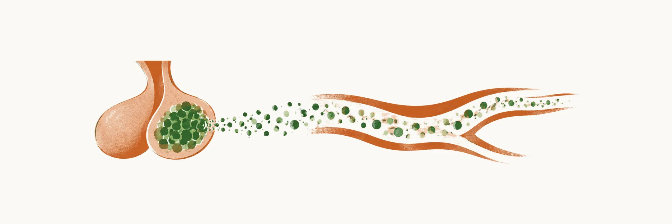
Acromegaly is caused by the excessive secretion of growth hormone (GH). Where that GH comes from, together with the type of adenoma and the molecular changes inside it, explains the behavior of the disease.
How acromegaly arises
Acromegaly is usually caused by an adenoma of the somatotrope cells, or GH-producing cells, of the anterior pituitary gland, but it may arise from other extra-pituitary lesions. GH-producing adenomas are benign lesions that arise from Pit-1 lineage cells of the anterior pituitary gland.
In more than 99% of the cases this excess secretion of GH is caused by the benign pituitary adenoma, and very rarely by:
- pituitary carcinomas
- ectopic secretion of GH by neuroendocrine tumors, namely lung, pancreatic and ovarian NETs
- excessive hypothalamic GHRH secretion, most often from chest and abdominal carcinoids
- iatrogenic injection of GH
Other causes and pseudoacromegaly
Not every picture of GH excess comes from a somatotrope adenoma, and not every picture of acral enlargement comes from GH excess at all.
| Pathology Associated with Acromegaly | |
|---|---|
| Pituitary adenoma | Densely granulated somatotroph adenoma; sparsely granulated somatotroph adenoma; mixed cell somatotroph and lactotroph adenoma; mammosomatotroph (monohormonal Pit-1 lineage) adenoma; acidophil stem cell adenoma; plurihormonal adenoma; poorly differentiated Pit-1 lineage tumor; pituitary hyperplasia; pituitary carcinoma; rare: ectopic pituitary adenomas identified in sphenoid sinus or parapharyngeal tissue |
| Ectopic hormone secretion | Central: hypothalamic tumors, ganglioneuroma. Peripheral: bronchial carcinoid, small cell lung cancer, adrenal tumor, pancreatic neuroendocrine tumor |
| Exogenous GH replacement or abuse | |
| Pseudoacromegaly | Pachydermoperiostosis; IGF signaling pathway diseases; severe insulin resistance |
Adenomas of the anterior pituitary
Pituitary adenomas are benign tumors that are derived from one of the five cell types of the anterior pituitary gland, so there are five main forms of adenoma, named after the cell they come from. The five cell types are the somatotrope (GH), the lactotrope (prolactin), the thyrotrope (TSH), the corticotrope (ACTH) and the gonadotrope (FSH and LH) cells. Acromegaly belongs to the somatotrope lineage, the GH-producing cells. Somatotrope adenomas make up about 10–15% of pituitary adenomas. Lactotrope adenomas are the most common form of functioning pituitary adenoma, with a prevalence of 50–60% of all functioning pituitary adenomas, and a further 15–35% of pituitary adenomas are non-functioning.
In the table, PRL is prolactin, ER the estrogen receptor, a-SU the alpha subunit, NOS not otherwise specified, and b-TSH, b-FSH and b-LH the beta subunits of TSH, FSH and LH. Adenomas are separated by lineage, transcription factors, the hormones they produce and their cytokeratin pattern:
| Adenoma type | Transcription Factors | Hormones | Cytokeratin |
|---|---|---|---|
| GH-producing adenomas | |||
| Densely granulated somatotroph adenoma | Pit-1 | GH, a-SU | diffuse |
| Sparsely granulated somatotroph adenoma | Pit-1 | GH | dot-like |
| Mammosomatotroph adenoma | Pit-1, ER | GH, PRL, a-SU | diffuse |
| Mixed somatotroph and lactotroph adenoma | Pit-1, ER | GH, PRL, a-SU | diffuse |
| PRL-producing adenomas | |||
| Sparsely granulated lactotroph adenoma | Pit-1, ER | PRL (Golgi) | diffuse |
| Densely granulated lactotroph adenoma | Pit-1, ER | PRL (diffuse) | diffuse |
| Acidophil stem-cell adenoma | Pit-1, ER | PRL (diffuse), GH | rare dot-like |
| TSH-producing adenoma | |||
| Thyrotroph adenoma | Pit-1, GATA-2 | b-TSH, a-SU | diffuse |
| ACTH-producing adenomas | |||
| Densely granulated corticotroph adenoma | Tpit | ACTH | diffuse |
| Sparsely granulated corticotroph adenoma | Tpit | ACTH | diffuse |
| Crooke’s cell adenoma | Tpit | ACTH | ring-like |
| Gonadotropin-producing adenoma | |||
| Gonadotroph adenoma | SF-1, GATA-2, ER | b-FSH, b-LH, a-SU | diffuse |
| Plurihormonal adenomas | |||
| Silent type III adenoma | Pit-1 (?), ER | multiple | diffuse |
| Unusual plurihormonal adenoma (NOS) | multiple | multiple | n/a |
| Hormone-negative adenoma | |||
| Null cell adenoma | none | none | diffuse |
Subtypes of somatotrope adenoma
Somatotrope adenomas are divided microscopically by how densely their cells are granulated, and that pattern predicts both the hormone output and the response to treatment.
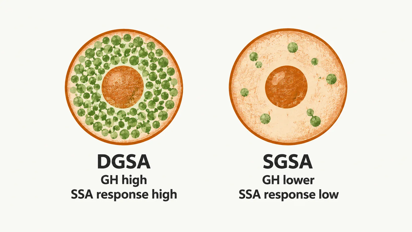
- Densely granulated somatotrope adenoma (DGSA) is the most common form. It is more common at older ages and is characterised by high secretion of GH, low proliferation and invasiveness, the GNAS mutation as its most common molecular change, and high responsiveness to somatostatin analogue (SSA) treatment.
- Sparsely granulated somatotrope adenoma (SGSA) is the second most common form. It is more prevalent in younger adults and in females, and it is characterised by a lower level of GH secretion, more proliferation and invasiveness, a lesser amount of GH positivity on staining, and lesser responsiveness to SSA treatment. A mixed-pattern adenoma, made up of both SGSA and DGSA, is also possible.
- Mammosomatotrope adenoma arises from a single Pit-1 lineage yet is positive for both GH and PRL. It is more common in younger adults, and because the prolactin it produces brings patients to medical attention earlier, it is usually diagnosed while still small and it is highly responsive to SSA treatment.
- Mixed somatotrope and lactotrope adenoma contains two cell populations and produces both hormones.
When an adenoma secretes both GH and prolactin, the clinical manifestation is sometimes dominated by the prolactin excess, and the acromegaly is not that evident to detect.
Molecular alterations in somatotrope adenomas
Several genes are altered in somatotrope adenomas; some bear on how the tumor behaves, others on how it responds to somatostatin analogue (SSA) treatment.
GNAS is the only recurrent genetic alteration in somatotrope adenomas. It encodes the Gsα portion of the G protein-coupled receptor (GPCR), and it is mutated in 15–58% of somatotrope adenomas, leading to constitutive activation of adenylate cyclase (AC). GPR101 acts on the same pathway. In patients with early-onset gigantism and GH hypersecretion there is a heritable microduplication of the X chromosome at Xq26.3, called X-linked acrogigantism (X-LAG), and the expression of Gpr101 mRNA in these patients is increased 1000-fold. Gpr101 is an orphan GPCR that is bound to Gsα and increases the activity of AC.
Two further groups of genes relate to the signals that control the somatotrope. Ghrelin is a growth factor that acts on the pituitary gland, and GHSR is the gene responsible for expression of the ghrelin receptor. For the somatostatin receptor genes, the pattern of expression differs between the subtypes of somatotrope adenoma and is highly influenced by SSA treatment: DGSA express more SSTR2 (somatostatin receptor type 2), while SGSA express mainly SSTR5, and the higher the expression of SSTR2, the higher the responsiveness to SSA treatment.
Aryl hydrocarbon interacting protein (AIP) gene. AIP mutations are mostly associated with somatotrope adenomas. The mutation is a non-sense mutation, so AIP products are believed to be onco-suppressor products, although information about them is still very limited. A germline AIP mutation is relatively common among young adults with acromegaly (5–14%), and it is associated with larger, more invasive adenomas with more potential for recurrence. Patients with an AIP mutation are relatively resistant to SSA treatment, whereas SSA treatment in patients without the mutation leads to upregulation of AIP expression.
Genetic and familial conditions
There are some genetic and familial conditions in which acromegaly can occur. Several of them rest on genes already met above: GPR101 in X-LAG, AIP in familial isolated pituitary adenoma, and GNAS in McCune–Albright syndrome. The others are the multiple endocrine neoplasia syndromes (MEN1 and MEN4), Carney complex, SDH (succinate dehydrogenase) mutations and neurofibromatosis.
| Genetic condition | Gene implicated | Inheritance | Clinical features |
|---|---|---|---|
| X-LAG | GPR101 | X-linked dominant or sporadic | Mostly females; age <5 years; hyperprolactinemia at diagnosis; can be de-novo and family history may be absent |
| Familial isolated pituitary adenoma | AIP | Autosomal dominant or sporadic | Younger males; higher GH levels; poor response to SSA therapy |
| MEN1 | MEN1 | Autosomal dominant or sporadic | Presence of primary hyperparathyroidism, pancreatic disease along with pituitary adenoma |
| MEN4 | CDKN1B | Autosomal dominant | Presence of pituitary and parathyroid neoplasms with pheochromocytomas, thyroid and other tumors |
| Carney complex | PRKAR1A | Autosomal dominant | Skin pigmentation, atrial myxoma, GH or PRL excess, Cushing’s due to primary pigmented nodular adrenocortical disease; somatotroph hyperplasia or multifocal adenoma |
| McCune–Albright | GNAS | Somatic mosaicism; not inherited | Average age of diagnosis 23 years; acromegaly seen in 20–30% of cases; hyperprolactinemia present at diagnosis; café-au-lait spots; peripheral precocious puberty; somatotroph hyperplasia |
| SDH mutation | SDHx | Autosomal dominant or sporadic | Phaeochromocytoma, paraganglioma and primary hyperparathyroidism present |
| Neurofibromatosis | NF1 | Autosomal dominant | Few case reports of children and adolescents with optic glioma; adenoma rarely reported; mechanisms unclear |