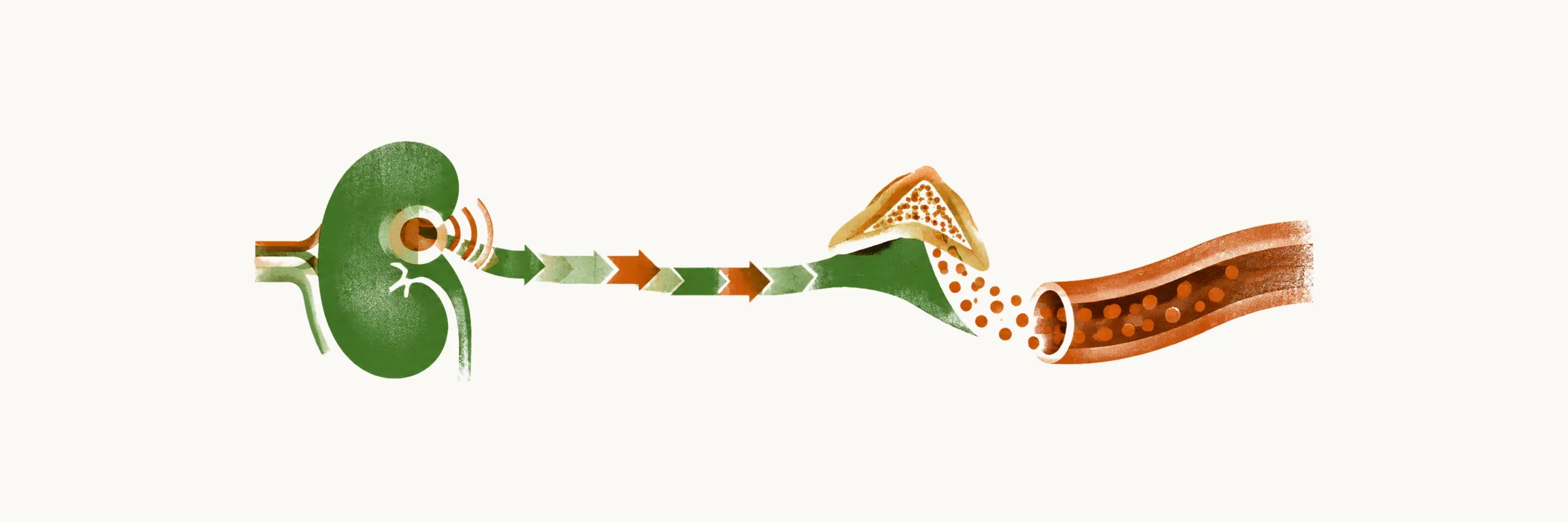
Aldosterone, the mineralocorticoid made by the zona glomerulosa of the adrenal cortex, is controlled mainly by the renin-angiotensin-aldosterone system, which links the kidney’s sense of perfusion to sodium retention and blood pressure, and the final step is an electrical event inside the zona glomerulosa cell.
How the macula densa sets renin release
The macula densa of the kidney modulates the secretion of renin according to the concentration of the chloride ion reaching it, and renin is the molecule that is the major controller of the renin-angiotensin-aldosterone system (RAAS). When the concentration of chloride reaching the macula densa falls, the juxtaglomerular cells that sit beside it in the wall of the afferent arteriole are activated to release renin into the systemic circulation. This mechanism is really a sensor of the perfusion of the kidney: the more hypoperfusion there is to the renal system, the lower the chloride reaching the macula densa. In hyperperfusion or hypertension, on the other hand, a higher level of chloride reaches the macula densa, and the release of renin is inhibited.
From renin to angiotensin II
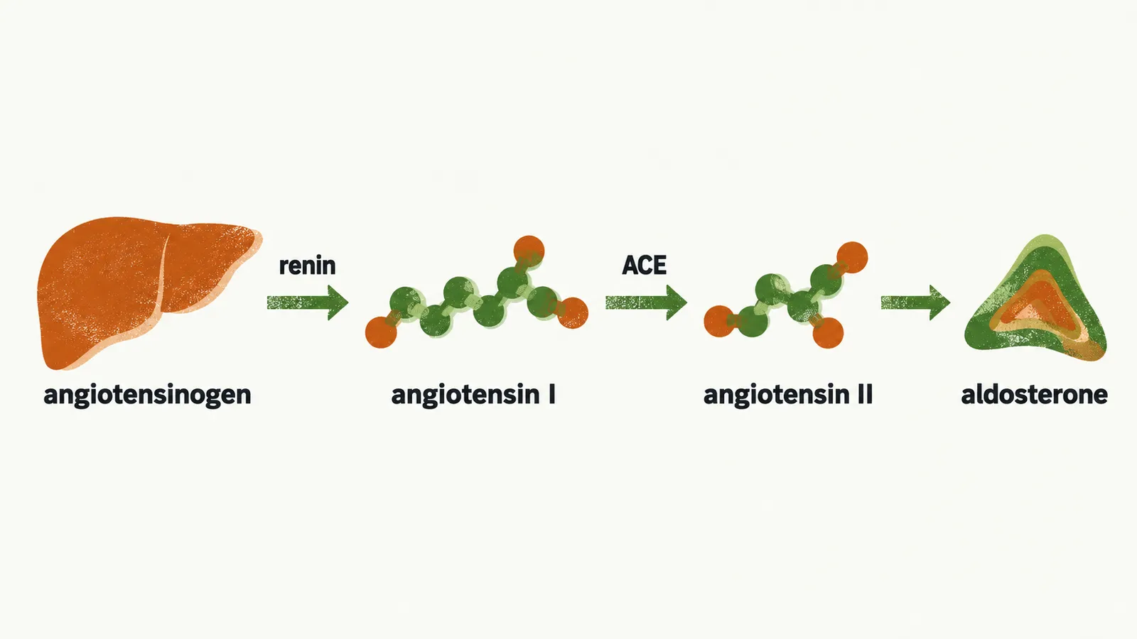
This chloride feedback is how the RAAS adjusts to hypotension physiologically. By activation of this feedback, angiotensinogen, which is made by the liver, is converted to angiotensin I thanks to the activity of renin. After the formation of angiotensin I, it is converted to angiotensin II by angiotensin-converting enzyme (ACE).
What angiotensin II does
The final physiological effect is exerted by angiotensin II, which acts in four ways:
- direct vasoconstriction;
- stimulating release of antidiuretic hormone (ADH), which increases water reabsorption and so increases intravascular volume and pressure;
- augmentation of sodium reabsorption by the proximal convoluted tubule (PCT), which increases intravascular water and sodium and so increases pressure;
- stimulating production of aldosterone by the zona glomerulosa of the adrenal glands, which increases sodium reabsorption by the distal convoluted tubule (DCT) and so increases pressure.
The first three actions act on vessels, water balance and the proximal tubule, whereas the fourth is the link to the adrenal cortex.
Other regulators of aldosterone
Renin, through angiotensin II, is one of the most important biological signals for the secretion of aldosterone, but it is not the only one. A rise in the plasma potassium concentration, or hyperkalaemia, stimulates aldosterone release directly, and this is the signal that couples aldosterone to potassium balance. ACTH also stimulates aldosterone, but its effect is very low and transient, so it does not set the day-to-day level of secretion.
How the zona glomerulosa cell secretes aldosterone
The zona glomerulosa senses potassium through its resting membrane potential. At rest, the cell is held hyperpolarised by background potassium channels that let potassium leak out of the cell. When the potassium concentration outside the cell rises, the gradient that drives that leak falls, so potassium efflux decreases and the membrane depolarises. The depolarisation opens voltage-gated calcium channels, chiefly of the T type, and the calcium that enters activates calcium-calmodulin-dependent kinases, which drive the expression of aldosterone synthase, or CYP11B2. A mutation that reduces the potassium current does the same thing in the absence of hyperkalaemia: the cell stays depolarised and aldosterone synthase is switched on. Angiotensin II converges on the same endpoint, because its receptor also raises intracellular calcium, so the two main controllers of aldosterone both act through calcium and CYP11B2.
Aldosterone is therefore controlled by renin, potassium and angiotensin II rather than mainly by ACTH, so its control loop is separate from that of cortisol. Cortisol has a different course after secretion: it is carried in the blood and switched on or off by tissues before it acts.