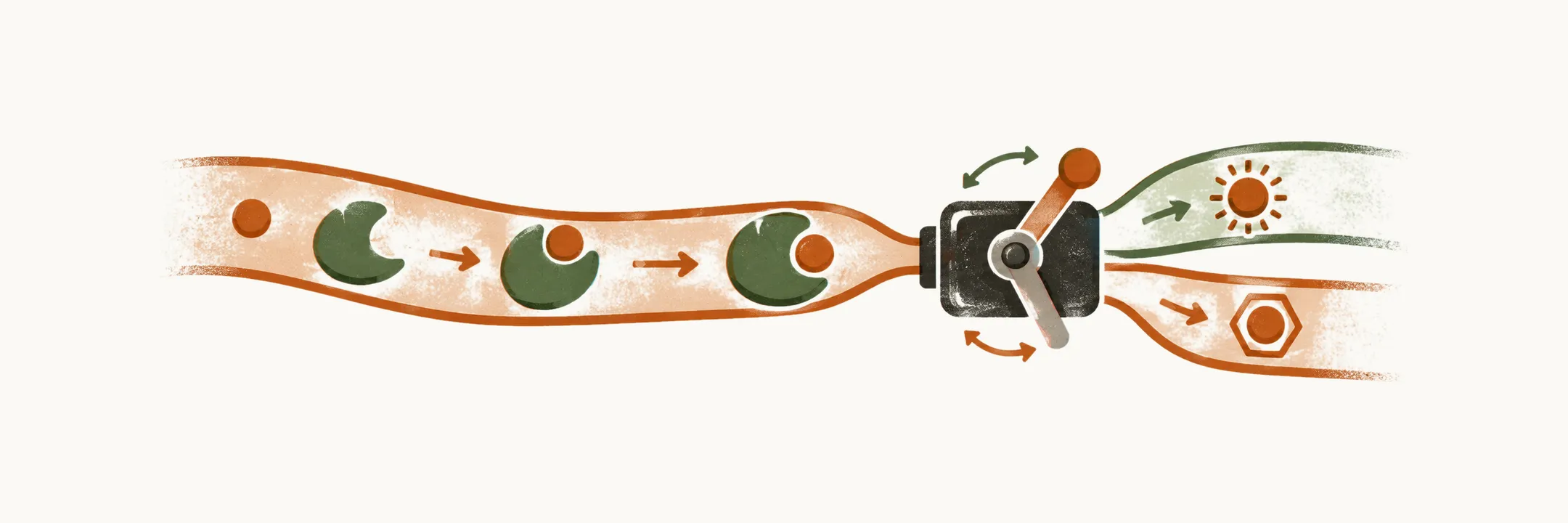
Circulating cortisol is not the same as active cortisol everywhere in the body: the hormone travels mostly bound to a carrier protein, and individual tissues switch it on or off before it reaches the glucocorticoid receptor that carries out its effects.
Cortisol and cortisone
The activated form of the glucocorticoid is cortisol, while the inactivated form is cortisone. The two are interconverted in tissues, and which form predominates depends on the enzymes a given tissue carries. Corticosterone, the glucocorticoid of the mineralocorticoid pathway, also circulates and has weak glucocorticoid activity of its own. It matters most when cortisol synthesis itself is blocked: in 17α-hydroxylase deficiency, for example, cortisol cannot be made, ACTH rises, and the precursors that would have led to cortisol are diverted into the mineralocorticoid pathway, so deoxycorticosterone and corticosterone accumulate. Because those steroids act on the mineralocorticoid receptor, the result is hypertension and hypokalaemia rather than a glucocorticoid effect.
Distribution in the blood
After production, cortisol is released into the bloodstream and is distributed among three fractions:
- the majority of the hormone is carried by cortisol-binding globulin (CBG);
- a minority of the hormone is carried by albumin;
- a very small portion travels as free hormone in the blood.
Only the free fraction can leave the vessel and enter cells, so a change in the amount of CBG changes the total measured cortisol without necessarily changing the free, active concentration.
Tissue conversion by 11-beta-HSD enzymes
In some tissues, cortisol, the activated form, is inactivated to cortisone by 11β-hydroxysteroid dehydrogenase type 2 (11β-HSD-2), mainly in the kidney, salivary gland and colon. In other tissues, such as adipose tissue, muscle, brain and liver, the inactivated form, cortisone, is converted back to cortisol by 11β-hydroxysteroid dehydrogenase type 1 (11β-HSD-1).
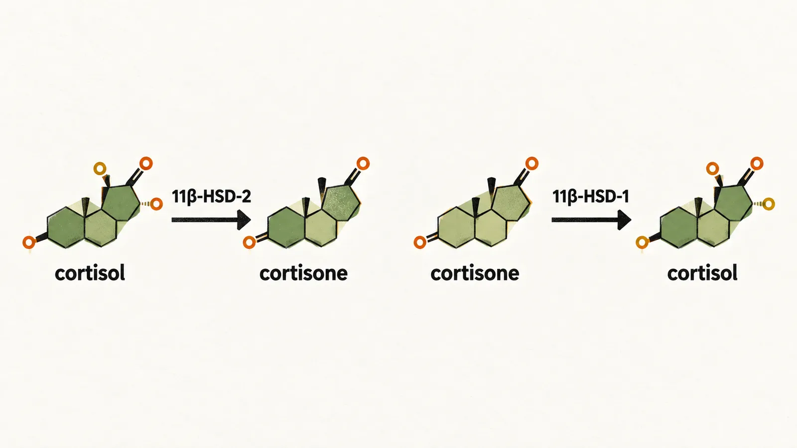
The two enzymes face in opposite directions, and their distribution decides how much active cortisol each tissue sees. In the kidney this matters especially, because the mineralocorticoid receptor there binds cortisol as readily as it binds aldosterone; 11β-HSD-2 inactivates cortisol before it can reach that receptor, so aldosterone keeps control of the site. Without the enzyme, cortisol would act as a mineralocorticoid.
Glucocorticoid receptor signalling
After cortisol enters a target cell, it attaches to the glucocorticoid receptor (GR). This causes heat shock proteins to dissociate from the receptor, the receptor and its ligand to dimerise, and the complex to enter the nucleus, where it activates or suppresses the synthesis of particular proteins. If the GR instead heterodimerises with NF-κB or AP-1, the complex acts as a suppressor of some genes, such as those for pro-inflammatory proteins.
Beyond this handling in the blood and tissues, the amount of cortisol available depends on the axis that drives its production, and that dependence is what makes adrenal function testable.