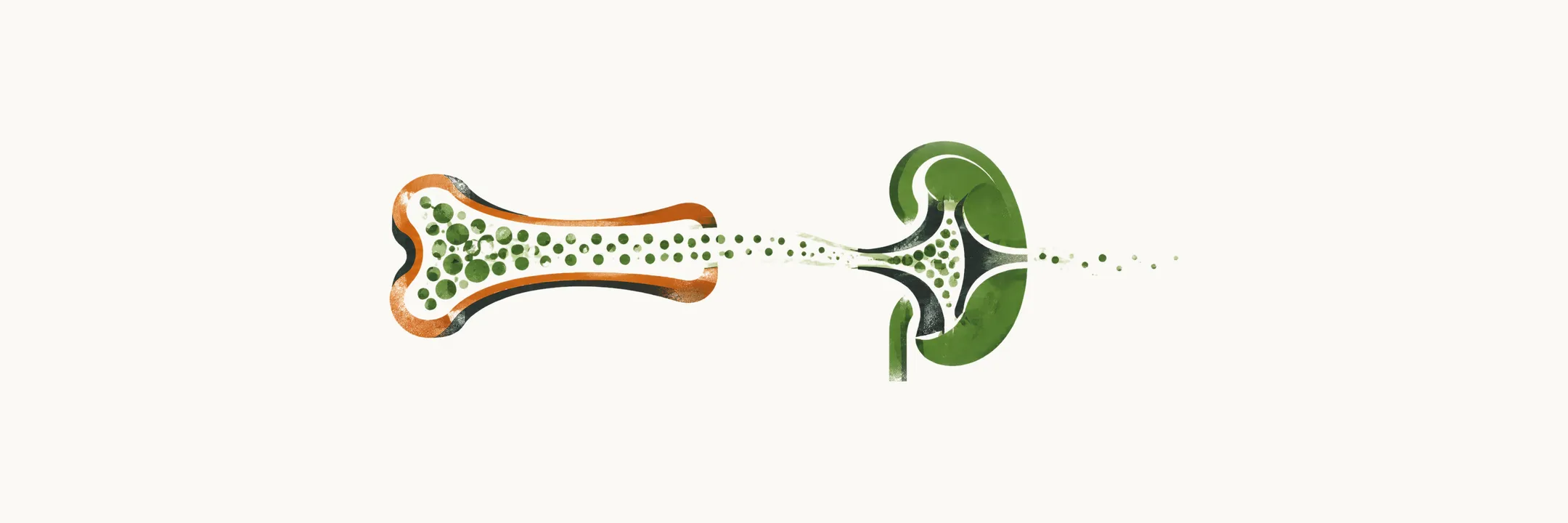
Calcium balance describes how much calcium the body holds, where it is held, and how it is taken in and lost.
Where the body’s calcium is
The total pool of calcium differs between healthy adult men and women: about 1300 g in men and about 1000 g in women. More than 99% of this pool is in the bones and teeth; the small remainder, in cells and in the extracellular fluid, is the part that carries the signal and sets the plasma level.
Intracellular calcium and calmodulin
The calcium concentration inside a cell is kept far below the concentration outside it: the intracellular calcium concentration stays between 100 nM and 1 microM, while the extracellular fluid is in the millimolar range. That steep gradient is the point of the arrangement, because calcium is used as a signal, and a small entry of calcium into the cytosol is a large relative change there, which is what allows it to trigger processes such as muscle contraction and secretion. Calcium exerts most of its effects inside the cell through calmodulin, a calcium-binding protein expressed throughout the body.
The plasma fractions
Outside the cells, the extracellular fluid calcium concentration is kept in a narrow range, 8.5-10.4 mg/dl or 2.1 to 2.6 mM, but the calcium in the extracellular fluid is not all in the same state. Three fractions make up the total:
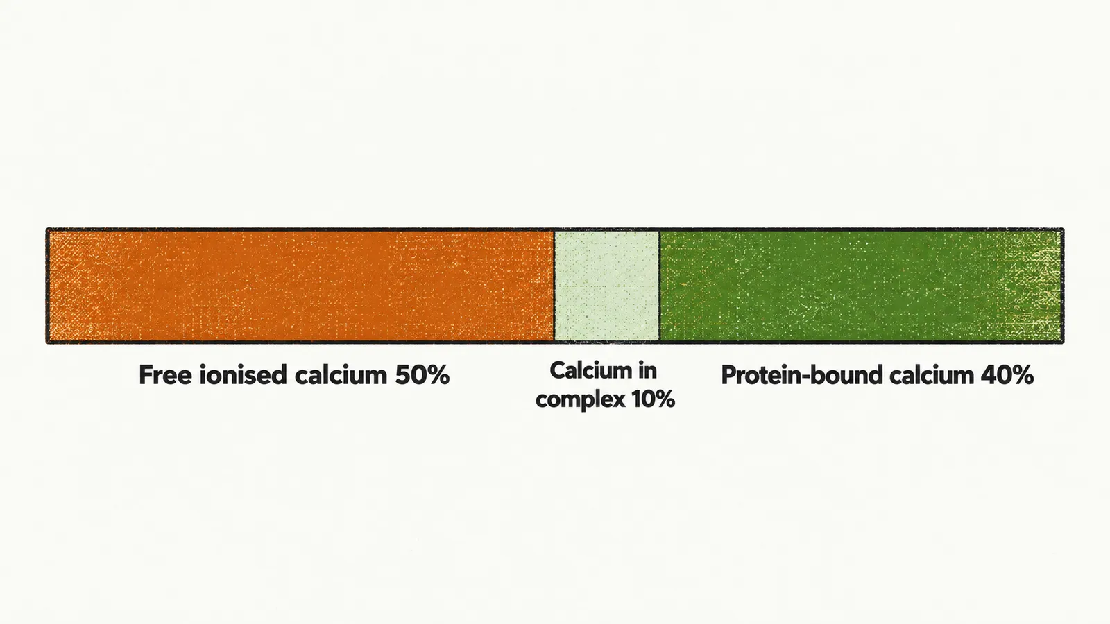
- Free ionised calcium — 50% of the 2.5 mM. This is the fraction the tissues respond to, and together with the calcium in complex it makes up the diffusible calcium, which is 1.5 mM.
- Calcium in complex — 10% of the 2.5 mM, bound to anions; the anionic part is mostly citrate or phosphate ions.
- Protein-bound calcium — 40% of the 2.5 mM, attached to plasma proteins; about 90% of the calcium attached to protein is attached to albumin.
Because most of the protein-bound share travels on albumin, a fall in albumin lowers the measured calcium without lowering the ionised fraction the tissues actually see. In physiological conditions, at a blood pH of 7.4, each 1 g/dl of albumin carries 0.8 mg/dl of calcium. To estimate the “adjusted” calcium level for protein binding, the following formula is used:
The same relationship runs in reverse: each 1 g/dl reduction in albumin reduces the extracellular fluid calcium concentration by 0.8 mg/dl.
Intestinal absorption
The only route of calcium absorption in the body is through the small intestine, and it happens in two forms:
- Active vitamin-D dependent calcium absorption, which relies on calcitriol, the active form of vitamin D. This process prevails over the passive one when the total intake of calcium is not adequate. Its location is the proximal duodenum.
- Passive calcium absorption, which happens when total intake of calcium is adequate or even high. Its location is the ileum or jejunum.
Calcium absorption in the intestine is less efficient in some conditions, such as in elderly people and with the use of some drugs, notably glucocorticoids and phenytoin.
Daily balance
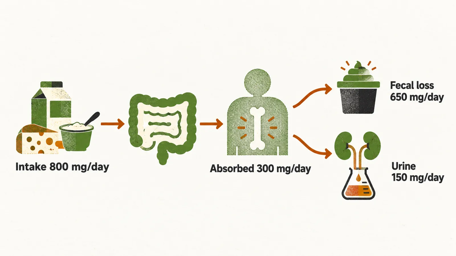
In normal physiological conditions, the amount of absorbed calcium equals the amount lost from the gastrointestinal tract (GIT) and kidney; this amount is mostly equal to 300 mg/day. Most adults take about 800 mg/day of calcium, of which 300 mg is absorbed. There is a fecal loss of 650 mg of calcium, of which 500 mg is from the diet and 150 mg is from the physiological loss of calcium in the GIT. In the US, more than 75% of the daily need for calcium intake is taken from milk and dairy products.
Renal handling
The kidney is the other route of loss. Every day about 9 g of calcium is filtered by the kidneys; more than 98% of it is reabsorbed, and about 150 mg/day is lost through the urine. Two parts of the kidney do the reabsorbing:
- the loop of Henle, by a passive mechanism that carries calcium along with sodium;
- the distal convoluted tubule (DCT), by an active mechanism governed by PTH, calcitonin and vitamin D.
Because the passive step in the loop of Henle follows sodium, the daily loss of calcium depends on what happens to sodium and water as well as on the hormones:
- the PTH level, which raises reabsorption in the distal tubule;
- daily sodium intake: the higher the sodium intake, the higher the calcium excretion;
- dietary protein intake: the calcium reabsorption function of the renal tubules depends heavily on proteins containing sulfur-containing amino acids;
- use of diuretics: loop diuretics increase the daily loss of calcium, while thiazide diuretics decrease it.
Gut and kidney therefore set how much calcium enters and leaves the body, and hormones adjust both steps. Phosphate moves through the same organs, is stored in the same bone, and is under some of the same hormonal controls, so it is the companion ion to understand before turning to the hormones themselves.