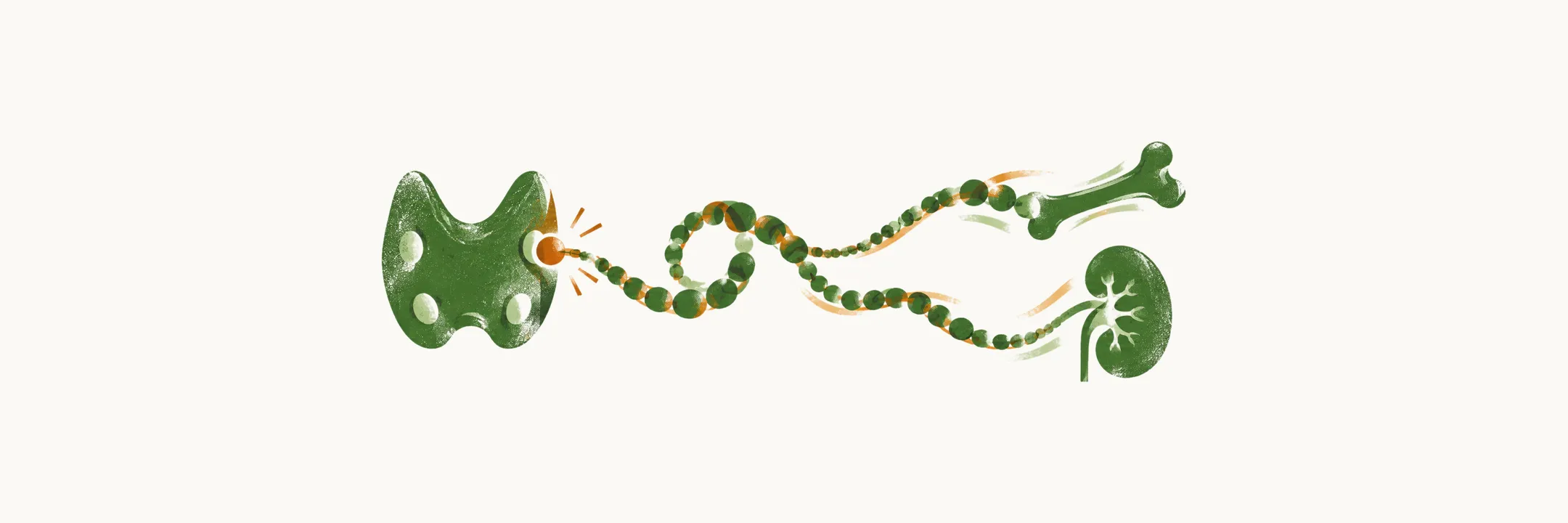
Parathyroid hormone (PTH) is the hormone of the parathyroid glands and the principal regulator of plasma calcium. The main hormonal factors that control the homeostasis of calcium and phosphate ions are PTH, fibroblast growth factor 23 (FGF23) and calcitriol, the active form of vitamin D. They act on the same main organs — bone, kidney and intestine — and PTH is the one that responds to calcium within minutes.
Synthesis
PTH is produced as a molecule of 115 amino acids called preproparathyroid hormone. It is cleaved by the ER (endoplasmic reticulum) to a 90-amino-acid proparathyroid hormone molecule, and then the removal of 6 amino acids in the Golgi produces PTH: a single polypeptide chain of 84 amino acids, whose first 34 amino acids from the N-terminal side carry the biological activity; the fragment PTH(1-34) is the shortest one that retains essentially complete activity.
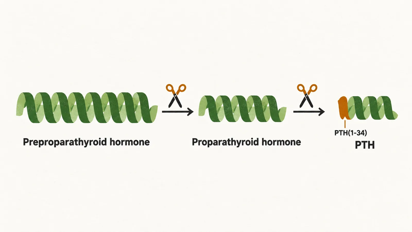
Changes at the two ends of the chain alter that activity. Studies show that if the 8th and 18th methionine are oxidised, the physiological activity is reduced. Derivatives that lack the first and second amino acids attach to the receptor but do not activate it, while derivatives that lack the first six amino acids inhibit PTH action. PTH has a half-life in the body of about 4 minutes and is mostly cleared by the liver and kidney.
Control of secretion
PTH secretion is based on the negative feedback of the calcium concentration in the plasma: when plasma calcium rises, PTH release falls, and when it falls, PTH is released. This happens thanks to specific plasma membrane calcium-sensing receptors that are GPCRs (G-protein-coupled receptors) bound to the G proteins Gi and Gq. These receptors are called CaSR.
In hypocalcemic conditions CaSR is not occupied by calcium, and Gi and Gq are not activated to inhibit the release of PTH, so PTH is released. When calcium occupies CaSR, two routes inhibit PTH release. The first runs through Gq: occupation of CaSR by the calcium ion activates Gq and increases PKC activity, and the resulting increase of intracellular calcium concentration leads to a reduction of PTH release. The second runs through Gi: Gi reduces the cAMP inside the parathyroid cells, and the release of PTH is inhibited by that route as well. Agents that increase the intracellular cAMP of the parathyroid cells increase the secretion of PTH, such as beta agonists and dopamine.
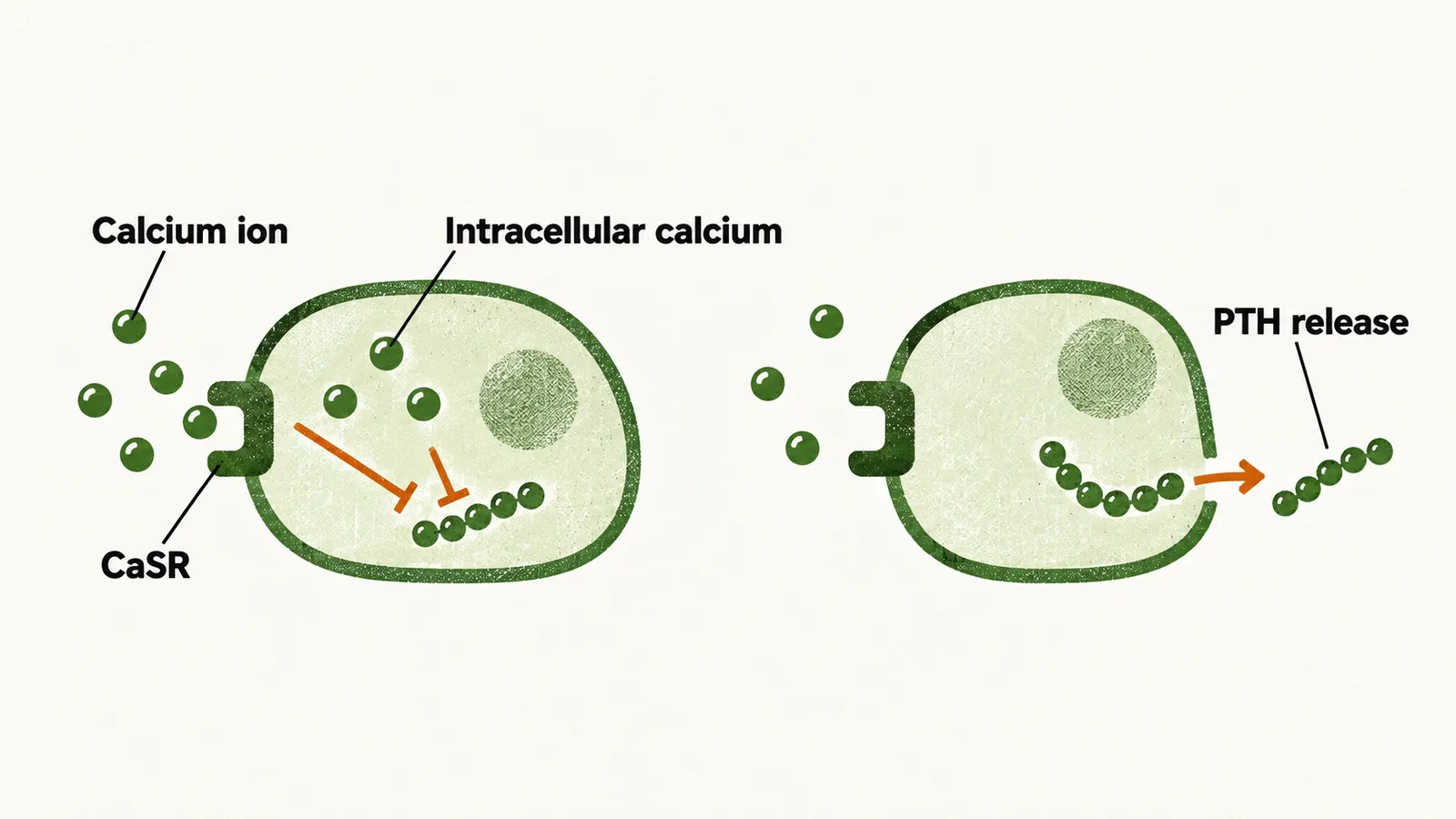
The Gq route is a physiological exception worth mentioning. In most cells of the body an increase of intracellular calcium is a stimulatory signal for secretion or expression, but in two types of cells an increase of intracellular calcium has an inhibitory effect on the secretion of a target product:
- the granular cells of the juxtaglomerular apparatus: an increase of intracellular calcium reduces the secretion of renin;
- parathyroid cells: an increase of intracellular calcium, promoted by occupation of CaSR by the calcium ion, reduces the release of PTH.
A loss-of-function mutation of the CaSR gene leads to familial hypocalciuric hypercalcemia type 1, or FHH-1.
The other, less important regulatory mechanisms of PTH secretion are:
- active vitamin D reduces PTH secretion;
- magnesium: hypermagnesemia inhibits PTH secretion, and hypomagnesemia also impairs it, so both extremes of magnesium disturb the axis.
Receptors
PTH acts through more than one receptor. There are at least three:
- type 1 PTH receptor, or PTH1R, which is responsible for the homeostatic and skeletal effects of PTH; PTH1R can also bind the PTH-related protein, or PTHrP
- type 2 PTHR, which is expressed on different organs such as cardiac tissue, pancreas, brain and placenta; it is activated only by PTH, not by PTHrP
- cPTHR, which binds the C-terminal end of the shortened PTH and is expressed on osteocytes
Actions
The physiological effects of PTH are calcium reabsorption in the kidney and phosphorus excretion in the kidney, together with calcium mobilization from bone and the stimulation of calcitriol production.
Calcium mobilization from the bone. The primary target cell of PTH in the bone is the osteoblast, which is the cell that then drives resorption by the osteoclast. PTH has both catabolic and anabolic effects on the bone: in the chronic release of PTH it is catabolic, causing bone loss, while intermittent release has an anabolic effect.
Stimulation of the conversion of the inactive to the active form of calcitriol in the kidney. This happens through the direct activation of 1α-hydroxylase of tubular cells. PTH also increases calcitriol production indirectly, through the hypophosphatemia caused by a high level of PTH, since hypophosphatemia itself increases calcitriol production.
Taken together, PTH raises plasma calcium and lowers plasma phosphate, and the fall in phosphate is what keeps calcium phosphate from precipitating in the tissues as calcium is mobilized.
Integrating the response
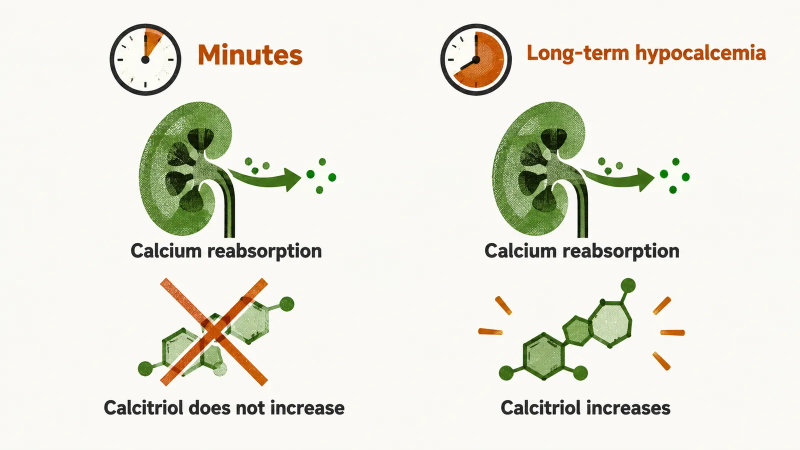
In a hypocalcemic condition, the changes in calcium, phosphate, calcitriol and PTH depend on the time course of the hypocalcemia:
- minute-to-minute calcium modulation: if the fall in calcium lasts only minutes, PTH only increases calcium reabsorption, which suffices for the compensation, and calcitriol does not need to increase in this circumstance
- long-term hypocalcemia: not only does the increased reabsorption of calcium happen, but calcitriol also increases
PTH therefore lowers phosphate and raises calcitriol. FGF23 is the hormone that also lowers phosphate but restrains calcitriol, and its release is itself driven by high PTH.